
Kimika Hara, contemporary hand embroiderer from Japan #womensart. Odra Noel. Photos: Scientists use microbes to make beautiful works of art in petri dishes. If you’ve ever found yourself buying clothes just because they’re cheap, or if shopping itself has become a form of entertainment for you, I’ve got a proposal: The next time you buy something, spend a whole lot on it.
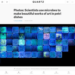
Enough that it makes you sweat a little. The point is to make you pause and ask yourself, “How much do I really want this?” In the US and much of the industrialized world, cheap clothes are everywhere. At any fast-fashion chain store, you’ll find piles upon piles of jeans that cost less than $20. The problem is, all that low-cost clothing is produced, sold, and finally discarded in mass quantities, which has serious consequences for the environment, the workers paid poorly to make them, and even the mental well-being of the people buying them. As a fashion reporter, I like clothes probably more than most. Two Pyramidals - Greg Dunn Design. 10 Award-Winning Microscope Images That Turn Science Into Art. Some of the most vibrant, beautiful photographs taken this year weren’t captured with a camera, but with a microscope.
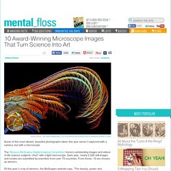
The Olympus BioScapes Digital Imaging Competition honors outstanding images and videos of life science subjects “shot” with a light microscope. Each year, nearly 2,500 still images and movies are submitted by scientists from over 70 countries. Psychedelic plants: The inner beauty of common species - Image 2. 12:29 03 November 2014 If you think "garlic" and "beauty" don’t belong in the same sentence, take a closer look at a magnified view of a tiny slice of the plant’s stem, captured by Rob Kesseler from the University of the Arts London (see below).
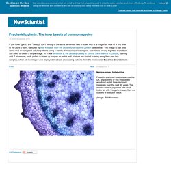
The image is part of a series that reveals plant cellular patterns using a variety of microscope techniques, sometimes piecing together more than 500 shots to create a single image. In a new exhibition at the Lethaby Gallery of Central Saint Martins in London, running until 7 November, each picture is blown up to span an entire wall. Visitors are invited to bring along their own tiny samples, which will be imaged and displayed in a book showcasing patterns from the microworld.
Janelia Research Campus. Zq issue 02, summer 2012 - Zygote QuarterlyZygote Quarterly. Home of David S. Goodsell. I recently completed a series of paintings showing the life cycle of HIV, as part of the book "The Future of HIV-1 Therapeutics: Resistance is Futile?
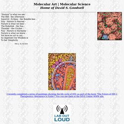
" You can see them at the HIVE Center WWW site. I got to talk a bit about my paintings with Brian Bartel and Dale Basler in an interview for "Lab Out Loud," a podcast presented by the National Science Teachers Association. Dan Klionsky directed a creative collaboration to explore the process of autophagy, which brought together choreographer and videographer Peter Sparling, composer Wendy Lee, and myself.
The result was a dance performance and a video: "Autophagy Suite: A Moving Diorama in Five Parts. " Click on the image above to see it! Drew Berry: DNA dances as imagination goes viral at White Night. Could not load plugins: File not found White Night 'virus' to infect State Library The State Library of Victoria is set to display a giant, 360 degree projection of animated human viruses as part of White Night. 14, 2014 Making something a billion times bigger - it sounds like a figure of speech or a wild exaggeration.

Yet it's exactly what Drew Berry is doing, and in the most unlikely, yet suitable, location. Drew Berry - Astonishing Molecular Machines. SUN Chloroplast E-book. Belle Cell by Angela Canada-Hopkins - Belle Cell Painting - Belle Cell Fine Art Prints and Posters for Sale. KANDINSKY + AUTOPHAGY = BLISS Background: Our... - Nature Graphics. Laura Splan - Doilies. Gallery: Karen Kamenetzky Fiber Art. The Fragile Cell: Karen Kamenetzky: Cellular art. Karen Kamenetzky is a textile artist making quilts inspired by microscopic/ cellular imagery.
" A kind of visual invented biology with textiles" Hypnotic Art Shows How Patterns Emerge From Randomness in Nature. Calgary art show explores stem-cell science. 12 January 2011by: | CBC News.
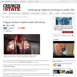
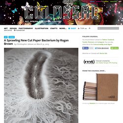
?articles. A Sprawling New Cut Paper Bacterium by Rogan Brown. Artist Rogan Brown (previously) recently completed work on this new cut paper sculpture titled Cut Microbe.
Institute of Astronomy. Glass artist Livvy Fink and poet Ezra Rubenstein, sparked by an ongoing collaboration between Cambridge Institute of Astronomy and Cancer Research UK, present a series of works exploring the relationship between macro-structures and micro-structures, and the sense of wonder they both provoke.
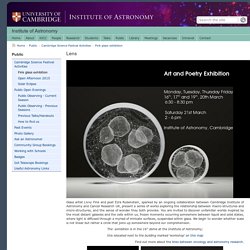
You are invited to discover unfamiliar worlds inspired by the most distant galaxies and the cells within us, frozen moments occurring somewhere between liquid and solid states, where light is diffused through a myriad of intricate surfaces, suspended within glass. We begin to wonder whether scale is not linear but rather a circle that joins up somewhere beyond our comprehension. The exhibition is in the 16" dome at the Institute of Astronomy; this islocated next to the building marked 'workshop' on this map. Benoit Mandelbrot Fractal Art Contest 2009 ~~ Entry: Cells Alive. Cells pictures on VisualizeUs.
Like Rob Kesseler found by Feya on oct 22, 2013 Like Rob Kesseler found by Feya on oct 22, 2013 Like 5 The Wondrous Beauty Of Microscopic Plant Seeds Co.Design business + design found by Feya on oct 22, 2013 Buy pictures with 20% off using the code: VISUALIZE20.
Microscopesandmonsters. This post takes me about 12 kilometres further north from the Durham coastal location that I wrote about in County Durham’s Tropical Seashore. I am in Seaham, on the top of cliffs made of the same Permian limestone that I found at Blackhall. Just beyond the Saxon church visible in some of the pictures is Seaham Hall Hotel, which has a very tenuous link with Lord Byron (he married the owner’s daughter; the marriage only lasted about a year before he ran off with his half-sister) and, more importantly, is now home to County Durham’s only Michelin-starred restaurant.
But nineteenth century literary scandal and gastronomy are not the reasons for my visit today. Microscopesandmonsters. My sampling trip to Upper Teesdale in search of desmids (see “Hunting for desmids in Upper Teesdale”) has now yielded another picture, this time figurative rather than semi-abstract. I have tried to depict the world inside a Sphagnum bog so have shown two desmids underneath a canopy of Sphagnum leaves.
The Sphagnum leaves have a characteristic structure, with chlorophyllose cells alongside water-filled “hyaline” cells. The desmids live, in effect, inside a glass-roofed conservatory although I have probably conveyed an overly bright impression of the subaquatic world of the bog. The reality is that the slow decay of Sphagnum yields brown humic materials that create an altogether murkier environment. The microscopic world of an Upper Teesdale Sphagnum bog, with desmids living in the space underneath Sphagnum leaves. Is this the first microscopic landscape painting? A few years ago I read Kenneth Clark’s book Landscape into Art, written in 1949 but which is still an excellent introduction to the history of landscape painting.
Timeless inner beauty… - AoB Blog. Image: P. Cuttings’ personal archive. When trying to appreciate something, it’s often remarked that it is the ‘inner beauty’ that’s important. In which case the plant cell biologists who probe the details within cells (and often illuminate them in all their glorious pin-point precision and fluorescent beauty with immunofluorescent techniques*) must not only, as scientists, be seekers of truth (for is it not writ, in scientia veritas?) Art Under the Microscope: Cell Press. Five questions with scientist-artist Deb Gumucio. It may not look like it, but the University of Michigan Center for Organogenesis, with its dissecting and transmission electron microscopes, doubles as an art studio. The center's director and founder, Deb Gumucio, is part of a collective of about 100 scientists-turned-artists who work at the Ann Arbor facility.
The research scientists at the center, who study the intricacies of cells and animal tissue to try and figure out why and how certain diseases occur, take pictures of their work. Gumucio and Sue O'Shea, director of human embryonic stem cell research, take submitted photographs of material that otherwise would be thrown away, and digitally enhance their tone and composition. Cell. Cell. The Micropolitan Museum.