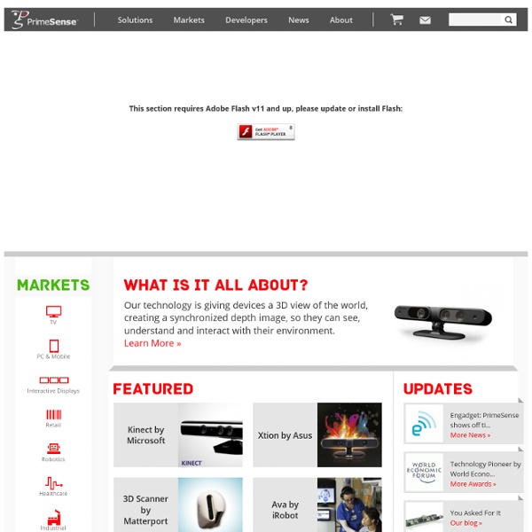


PrimeSense reveals Capri, 'world's smallest' 3D sensor PrimeSense™ Unveils Capri, World's Smallest 3D Sensing Device at CES 2013 TEL AVIV, Israel, Dec. 11, 2012 /PRNewswire/ -- PrimeSense™ ( the leader in Natural Interaction and 3D sensing solutions, today announced the launch of its next generation embedded 3D sensor, Capri, demonstrating a revolutionary small form factor and low cost. PrimeSense will debut Capri as part of its World of 3D Sensing suite at the Renaissance Hotel in Las Vegas, January 8-11 at the 2013 International Consumer Electronics Show (CES). PrimeSense's breakthrough reference design utilizes Capri - PrimeSense's next-generation of depth acquisition System on Chip, with improved algorithms including multi-modal 3D sensing techniques. "Using cutting-edge technologies, our newest generation of sensors is robust, accurate and affordable," said Inon Beracha, CEO, PrimeSense.
Dale Phurrough Cycling 74 Max external using Microsoft Kinect SDK dp.kinect is an external which can be used within the Cycling ’74 Max development environment to control and receive data from your Microsoft Kinect. Setup and usage docs are available at EASY Kinect 3D Scanner! Hey Instructables community! In this instructable I will instruct you how to make a DIY 3d scanner using an XBOX-360 Kinect! This instructable is very easy as long as you are patient and follow my instructions in the video. Also, I explain how to correct and fix your image for a nice clean print on a 3D printer such as the UP.
Technology Inc. - Microchip’s New GestIC® Technology Enables Mobile-Friendly 3D Gesture Interfaces Images High-res Photo Available Through Flickr or Editorial Contact (feel free to publish): Download Hi-Res New Kinect Gets Closer to Your Body [Videos, Links] The new, svelte-looking Kinect. It’s not that it looks better, though, that matters: it’s that it sees better. Courtesy Microsoft. Virtual Fitting Room » Case studies For REAL statistics, REAL measures of performance and REAL opinions on the Fits.me Virtual Fitting Room, read the case studies below. There’s no need to take our word for it when there are others to speak for us. Henri Lloyd case study – October 2013 “The stats we’re getting are overwhelming evidence that our virtual fitting room….and as a result it has smashed the overall returns rate.”Graham Allen Click on image to download
CEVA Gesture Recognition solutions for mobile and home entertainment devices eyeSight is a leader in Touch Free Interfaces for digital devices. The company was established with the vision to revolutionize the way people interact with digital devices, to create an interaction which is both simple and intuitive. eyeSight’s solution is based on advanced image processing and machine vision algorithms, which analyze real time video input from common built-in cameras. This technology is designed for embedded platforms. therenect - A virtual Theremin for the Kinect Controller The Therenect is a virtual Theremin for the Kinect controller. It defines two virtual antenna points, which allow controlling the pitch and volume of a simple oscillator. The distance to these points can be adjusted by freely moving the hand in three dimensions or by reshaping the hand, which allows gestures that should be quite similar to playing an actual Theremin. This video was recorded prior to this release, an updated video introducing the improved features of the current version will follow soon. Configuration Oscillator: Theremin, Sinewave, Sawtooth, Squarewave Tonality: continuos mode or Chromatic, Ionian and Pentatonic scales MIDI: optionally send MIDI note on/off events to the selected device & channel Kinect: adjust the sensor camera angle AcknowledgmentsThis application has been created by Martin Kaltenbrunner at the Interface Culture Lab.
Explore Cornell - The 3D Body Scanner - Made-to-Measure Twenty-first century technologies are defining a new era of customized and mass-customized clothing. Worldwide, apparel firms are experimenting with economical strategies that individualize clothing for each customer by offering a variety of design and fit options. Large and small, Internet as well as bricks-and-mortar companies are now making clothing "just for you." Levi Strauss & Co. was the first large apparel company to offer mass customization when they introduced "Personal Pair" jeans, later marketed under the name "Original Spin", in selected Levi's stores. Consumers could customize their jeans by choosing from a selection of styles, fabrics, finishes, colors, leg-opening sizes, and inseam lengths. Individual measurements were taken by a salesperson.
PCs of the near future: Intel lays out next-gen plans LAS VEGAS--PCs on your coffee table, playing Monopoly. Super-thin ultrabooks. Voice and gestural computing. Intel showed these and more at their CES 2013 press conference. But does it add up to a firm control on the future of computing? Fourth-gen Intel Core processors aren't on their way immediately, but at this year's CES Intel was ready to demonstrate how its "Haswell" code-named chips will make Windows 8 devices of tomorrow even thinner and smaller than now ... if you're in need of that.
Support » Skanect 3D Scanning Software By Occipital Contact You can email us at skanect@occipital.com or get help from the Skanect community, in the skanect google group. Tutorials SixthSense - a wearable gestural interface (MIT Media Lab) 'SixthSense' is a wearable gestural interface that augments the physical world around us with digital information and lets us use natural hand gestures to interact with that information. We've evolved over millions of years to sense the world around us. When we encounter something, someone or some place, we use our five natural senses to perceive information about it; that information helps us make decisions and chose the right actions to take. But arguably the most useful information that can help us make the right decision is not naturally perceivable with our five senses, namely the data, information and knowledge that mankind has accumulated about everything and which is increasingly all available online.
PrimeSense’s depth acquisition is enabled by "light coding" technology. The process codes the scene with near-IR light, light that returns distorted depending upon where things are. The solution then uses a standard off-the-shelf CMOS image sensor to read the coded light back from the scene using various algorithms to triangulate and extract the 3D data. The product analyses scenery in 3 dimensions with software, so that devices can interact with users by userexperience Jan 23
PrimeSense System on a Chip (SoC) The CMOS image sensor works with the visible video sensor to enable the depth map provided by PrimeSense SoC’s Carmine (PS1080) and Capri (PS1200) to be merged with the color image. The SoCs perform a registration process so the color image (RGB) and depth (D) information is aligned properly.[8] The light coding infrared patterns are deciphered in order to produce a VGA size depth image of a scene. It delivers visible video, depth, and audio information in a synchronized fashion via the USB 2.0 interface. The SoC has minimal CPU requirements as all depth acquisition algorithms run on the SoC itself. by userexperience Jan 23