
Neural oscillation. Neural oscillation is rhythmic or repetitive neural activity in the central nervous system.
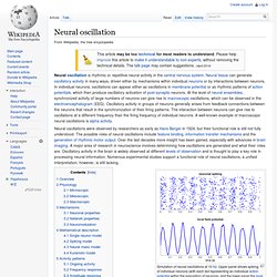
Neural tissue can generate oscillatory activity in many ways, driven either by mechanisms within individual neurons or by interactions between neurons. In individual neurons, oscillations can appear either as oscillations in membrane potential or as rhythmic patterns of action potentials, which then produce oscillatory activation of post-synaptic neurons. Mu wave. One second sample of an EEGalpha wave recording.
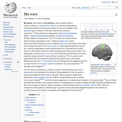
This wave occurs at frequencies similar to the mu wave, although the alpha wave is detected over a different part of the brain. Mu waves, also known as mu rhythms, comb or wicket rhythms, arciform rhythms, or sensorimotor rhythms, are synchronized patterns of electrical activity involving large numbers of neurons, probably of the pyramidal type, in the part of the brain that controls voluntary movement.[1] These patterns as measured by electroencephalography (EEG), magnetoencephalography (MEG), or electrocorticography (ECoG) repeat at a frequency of 7.5–12.5 Hz and are most prominent when the body is physically at rest.[1] Unlike the alpha wave, which occurs at a similar frequency over the resting visual cortex at the back of the scalp, the mu wave is found over the motor cortex, in a band approximately from ear to ear.
Delta wave. Theta rhythm. Example of an EEG theta wave The theta rhythm is an oscillatory pattern in electroencephalography(EEG) signals recorded either from inside the brain or from electrodes glued to the scalp.
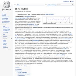
Two types of theta rhythm have been described. The "hippocampal theta rhythm" is a strong oscillation that can be observed in the hippocampus and other brain structures in numerous species of mammals including rodents, rabbits, dogs, cats, bats, and marsupials. "Cortical theta rhythms" are low-frequency components of scalp EEG, usually recorded from humans. In rats, the most frequently studied species, theta rhythmicity is easily observed in the hippocampus, but can also be detected in numerous other cortical and subcortical brain structures.
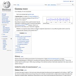
The function of the hippocampal theta rhythm is not clearly understood. Cortical theta rhythms observed in human scalp EEG are a different phenomenon, with no clear relationship to the hippocampus. Gamma wave. Gamma waves A gamma wave is a pattern of neural oscillation in humans with a frequency between 25 and 100 Hz,[1] though 40 Hz is typical.[2] According to a popular theory, gamma waves may be implicated in creating the unity of conscious perception (the binding problem).[3][4][5] However, there is no agreement on the theory; as a researcher suggests:
Beta wave. Beta wave, or beta rhythm, is the term used to designate the frequency range of human brain activity between 12.5 and 30 Hz (12.5 to 30 transitions or cycles per second).
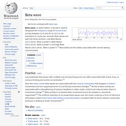
Beta waves are split into three sections: Low Beta Waves (12.5–16 Hz, "Beta 1 power"); Beta Waves (16.5–20 Hz, "Beta 2 power"); and High Beta Waves (20.5–28 Hz, "Beta 3 power").[1] Beta states are the states associated with normal waking consciousness. Function[edit] Low amplitude beta waves with multiple and varying frequencies are often associated with active, busy, or anxious thinking and active concentration.[2] Alpha wave. Alpha waves are neural oscillations in the frequency range of 7.5–12.5 Hz[1] arising from synchronous and coherent (in phase or constructive) electrical activity of thalamic pacemaker cells in humans.
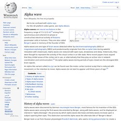
They are also called Berger's wave in memory of the founder of EEG. Alpha waves are one type of brain waves detected either by electroencephalography (EEG) or magnetoencephalography (MEG) and predominantly originate from the occipital lobe during wakeful relaxation with closed eyes. Alpha waves are reduced with open eyes, drowsiness and sleep. Historically, they were thought to represent the activity of the visual cortex in an idle state. Sleep spindle. A sleep spindle is a burst of oscillatory brain activity visible on an EEG that occurs during stage 2 sleep.
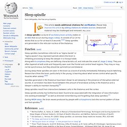
It consists of 12–14 Hz waves that occur for at least 0.5 seconds.[1][2] Sleep spindles are generated in the reticular nucleus of the thalamus. Function[edit] Sleep spindles (sometimes referred to as "sigma bands" or "sigma waves") may represent periods where the brain is inhibiting processing to keep the sleeper in a tranquil state. Along with K-complexes they are defining characteristics of, and indicate the onset of, stage 2 sleep. They are often tapered at both ends and frequently seen over the frontal and central head regions.
Sensorimotor rhythm. K-complex. A K-complex is an electroencephalography (EEG) waveform that occurs during stage 2 of NREM sleep.
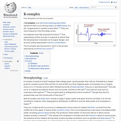
It is the "largest event in healthy human EEG".[1] They are more frequent in the first sleep cycles. K-complexes have two proposed functions:[1] first, suppressing cortical arousal in response to stimuli that the sleeping brain evaluates not to signal danger, and second, aiding sleep-based memory consolidation. The K-complex was discovered in 1937 in the private laboratories of Alfred Lee Loomis.[2] Neurophysiology[edit] K-complex consists of a brief negative high-voltage peak, usually greater than 100 µV, followed by a slower positive complex around 350 and 550 ms and at 900 ms a final negative peak.