
ReachMD - Reaching and Teaching Medical Professionals - XM Radio 245. Genes to Cognition Online. Free Medical Books. This man’s photo’s are AMAZING! I can’t wait till... Right upper lobe collapse. Right upper lobe collapse has distinctive features, and is usually easily identified on frontal chest radiographs; much more so than left upper lobe collapse.
For a general discussion please refer to the article on lobar collapse. Radiographic features. Focal Parenchymal Lung Lesions Showing a Potential of False-Positive and False-Negative Interpretations on Integrated PET/CT : American Journal of Roentgenology: Vol. 186, No. 3 (AJR) Inflammatory or Infectious Lung Lesions Simulating Lung Cancer with Increased 18F-FDG Uptake Active tuberculosis or tuberculoma, acute and chronic pneumonia, abscess, fungal infection, sarcoidosis, parasitic infestation, and pneumoconiosis are frequent causes of increased 18F-FDG uptake.

Benign Neoplasms with Increased 18F-FDG Uptake Simulating Malignancy Benign tumors such as sclerosing hemangioma, leiomyoma, and inflammatory pseudotumor may show increased 18F-FDG uptake. Lung Neoplasms with Reduced 18F-FDG Uptake Simulating Benignancy. How to interpret CT scans of your lung. Table of contents 1.
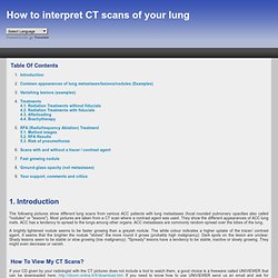
Introduction The following pictures show different lung scans from various ACC patients with lung metastases (focal rounded pulmonary opacities also called "nodules" or "lesions"). Most pictures are taken from a CT scan where a contrast agent was used. They show the different appearances of ACC lung mets. A brightly lightened nodule seems to be faster growing than a greyish nodule. How to view my CT scans? If your CD given by your radiologist with the CT pictures does not include a tool to watch them, a good choice is a freeware called UNIVIEWER that can be downloaded here: If you need to know how to use UNIVIEWER send us an email and ask for assistance. What do I see on the pictures? A CT scan makes mirror images. You are also able to see the different lobes. Chest Radiology. Chest xray. Basics of Chest X-ray Interpretation: A Programmed Study Acknowledgment is given to Leslie Muma, RN, MSN, NP for assistance in prepartion of this learning module.
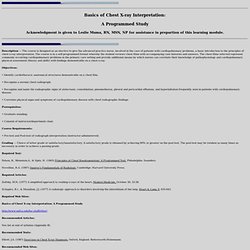
Description — The course is designed as an elective to give the advanced practice nurse, involved in the care of patients with cardiopulmonary problems, a basic introduction to the principles of chest x-ray interpretation. The course is in a self-programmed format whereby the student reviews chest films with accompanying case histories and answers. The chest films selected represent commonly occurring cardiopulmonary problems in the primary care setting and provide additional means by which nurses can correlate their knowledge of pathophysiology and cardiopulmonary physical assessment (theory and skills) with findings demonstrable on a chest x-ray. Objectives: • Identify cardiothoracic anatomical structures demonstrable on a chest film. Instant Anatomy - Thorax - Muscles - Attached to costal cartilages.
Kinesiology of the Shoulder and Scapula. Cerebro - neurociencia. Atlas of Human Anatomy in Cross Section: Section 5. Lower Thorax (Lungs) and Abdomen. Home | Search | About Us | FAQ | Reviews | Contact Us Atlas of Human Anatomy in Cross Section: Section 5.
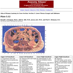
Lower Thorax (Lungs) and Abdomen Ronald A. Bergman, Ph.D., Adel K. Afifi, M.D., Jean J. This section passes through the third lumbar vertebra (25), its transverse process (34), spine, and superior articular process (30). On the left side, the transverse colon (47) is seen as smooth walled tubes, followed by loops of jejunum (44), which shows folds and villi of mucosa. Anatomy Dissection Videos - Free medical Video Lectures. Anatomy. Human Anatomy Course Human Anatomy UPPER LIMB 2 of 8. PPT - human anatomy Powerpoint Slide - Presentations.
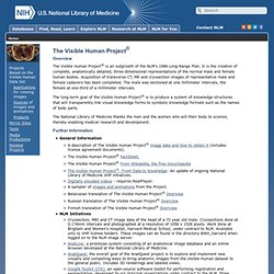
Biceps Brachii Muscle (Short Head) Radiographic Anatomy of the Skeleton: Lumbar Spine. Department of Neurobiology and Developmental Sciences - Muscles of the Upper Limb. Southern California Orthopedic Institute (SCOI) The two main bones of the shoulder are the humerus and the scapula (shoulder blade).
The joint cavity is cushioned by articular cartilage covering the head of the humerus and face of the glenoid. The scapula extends up and around the shoulder joint at the rear to form a roof called the acromion, and around the shoulder joint at the front to form the coracoid process. The National Library of Medicine's Visible Human Project. Overview The Visible Human Project® is an outgrowth of the NLM's 1986 Long-Range Plan.
It is the creation of complete, anatomically detailed, three-dimensional representations of the normal male and female human bodies. Acquisition of transverse CT, MR and cryosection images of representative male and female cadavers has been completed. The male was sectioned at one millimeter intervals, the female at one-third of a millimeter intervals. The long-term goal of the Visible Human Project® is to produce a system of knowledge structures that will transparently link visual knowledge forms to symbolic knowledge formats such as the names of body parts. The National Library of Medicine thanks the men and the women who will their body to science, thereby enabling medical research and development. Further Information. Workshop Anatomy for the Internet: international homepage. Atlas of Human Anatomy in Cross Section: Section 4. Upper Limb.
Home | Search | About Us | FAQ | Reviews | Contact Us Atlas of Human Anatomy in Cross Section: Section 4.
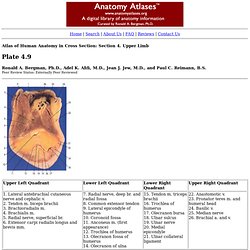
Upper Limb Ronald A. Bergman, Ph.D., Adel K. Afifi, M.D., Jean J. Atlas of Human Anatomy: Plate 13. Translated by: Ronald A.
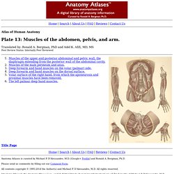
Bergman, PhD and Adel K. Afifi, MD, MS Peer Review Status: Internally Peer Reviewed Anatomy Atlases is curated by Michael P. D'Alessandro, M.D. [Google+ Profile] and Ronald A. Please send us comments by filling out our Comment Form. Nerves and Vessels to Superficial Layer of Muscles of the Back.
Medical Mnemonics .com: World's Database of Medical Mnemonics. Discover the Human Body: Interactive Anatomy Guide. GetBodySmart: Interactive Tutorials and Quizzes On Human Anatomy and Physiology. Basic Human Anatomy: Table of contents. LUMEN - Structure of the Human Body. List of Upper Limb Muscles.