
Crescendo-decrescendo murmur - definition of crescendo-decrescendo murmur in the Medical dictionary. Murmur /mur·mur/ (mur´mer) [L.] an auscultatory sound, particularly a periodic sound of short duration of cardiac or vascular origin. anemic murmur a cardiac murmur heard in anemia. aortic murmur one generated by blood flowing through a diseased aorta or aortic valve. arterial murmur one over an artery, sometimes aneurysmal and sometimes constricted.

Ventricular Ectopic Beats. Blood pressure. Blood pressure without further specification usually refers to the systemic arterial pressure measured at a person's upper arm and is a measure of the pressure in the brachial artery, the major artery in the upper arm.
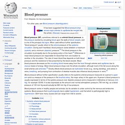
A person’s blood pressure is usually expressed in terms of the systolic pressure over diastolic pressure and is measured in millimetres of mercury (mm Hg), for example 120/80. It is also expressed as the amount over normal atmospheric pressure (760 mm Hg), so a blood pressure of 120 mm Hg would actually be 880 mm Hg of true pressure. Blood pressure varies in healthy people and animals, but its variation is under control by the nervous and endocrine systems. Blood pressure that is pathologically low is called hypotension, and that which is pathologically high is hypertension. Both have many causes and can range from mild to severe. Ectopic Beats: Description, Symptoms, Risks and Treatment. Posted by webmaster An Ectopic Beat or Cardiac ectopy is an irregular beat arising in the heart due to variations in the electrical conductance system of the heart. Ectopic beats are generally harmless and may occur without any particular cause.
Ectopic beats are common in adults and if it happens frequently, an examination should be done, even if no further treatment is prescribed. Ectopic Beats may either be. What Is Heart Ablation? Impedance cardiography. Impedance cardiography (ICG) is a plethysmography technique of using sensors to detect the properties of the blood flow in the thorax.

Introduction[edit] Impedance cardiography (ICG), also referred to as electrical impedance plethysmography (EIP), has been researched since the 1940s. NASA helped develop the technology in the 1960s.[1][2] The use of impedance cardiography in psychophysiological research was pioneered by the publication of an article by Miller and Horvath in 1978.[3] Subsequently, the recommendations of Miller and Horvath were confirmed by a standards group in 1990.[4] A comprehensive list of references is available at ICG Publications.
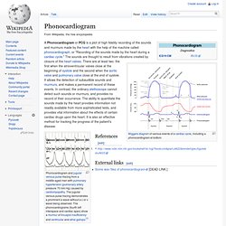
With ICG, the placement of four dual disposable sensors on the neck and chest are used to transmit and detect electrical and impedance changes in the thorax, which are used to measure and calculate hemodynamic parameters. How ICG Works[edit] Ballistocardiography. The ballistocardiograph (BCG) is a measure of ballistic forces on the heart.[1] Ballistocardiography is a technique for producing a graphical representation of repetitive motions of the human body arising from the sudden ejection of blood into the great vessels with each heart beat.[2] It is a vital sign in the 1-20 Hz frequency range which is caused by the mechanical movement of the heart and can be recorded by noninvasive methods from the surface of the body.

It was shown for the first time, after an extensive research work by Dr. Phonocardiogram. References[edit] External links[edit] Some wav files of phonocardiogram [DEAD LINK.]
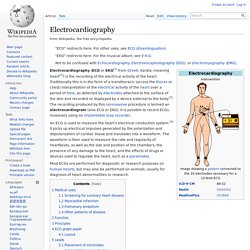
Echocardiography. Echocardiogram in the parasternal long-axis view, showing a measurement of the heart's left ventricle Echocardiogram, often referred to as a cardiac echo or simply an echo, is a sonogram of the heart.
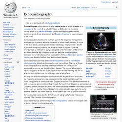
(It is not abbreviated as ECG, which in medicine usually refers to an electrocardiogram.) Echocardiography uses standard two-dimensional, three-dimensional, and Doppler ultrasound to create images of the heart. Echocardiography has become routinely used in the diagnosis, management, and follow-up of patients with any suspected or known heart diseases. It is one of the most widely used diagnostic tests in cardiology.
Echocardiography can help detect cardiomyopathies, such as hypertrophic cardiomyopathy, dilated cardiomyopathy, and many others. Not only can an echocardiogram create ultrasound images of heart structures, but it can also produce accurate assessment of the blood flowing through the heart, using pulsed or continuous wave Doppler ultrasound. Cardiotocography. The invasive fetal monitoring was invented by Doctors Alan Bradfield, Orvan Hess and Edward Hon.

A refined (antepartal, non-invasive, beat-to-beat) version (cardiotocograph) was later developed for Hewlett Packard by Dr. Konrad Hammacher. Photoplethysmogram. Representative PPG taken from an ear pulse oximeter.

Variation in amplitude are from Respiratory Induced Variation. Cardiography Systems. What Is Angina. Angina (an-JI-nuh or AN-juh-nuh) is chest pain or discomfort that occurs if an area of your heart muscle doesn't get enough oxygen-rich blood.
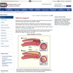
Angina may feel like pressure or squeezing in your chest. The pain also can occur in your shoulders, arms, neck, jaw, or back. Angina pain may even feel like indigestion. Angina isn't a disease; it's a symptom of an underlying heart problem. Electrocardiography. "EKG" redirects here.
For the musical album, see E·K·G. Angiocardiography. Angiocardiography is a technique for radiographic examination of the heart chambers and thoracic veins and arteries. A liquid radiocontrast agent, typically containing iodine, is injected into the bloodstream, then the tissues are examined using X-rays.[1] To avoid dilution, the radiopaque material is typically introduced with a catheter, a process known as selective angiocardiography.