
NINDS News Articles.
M16%20(CBT435)-Abdominal%20Emerg. The Role of the Vagus Nerve in Bulimia. Viva Las Vagus Nerve. A corny title – but a cool nerve.
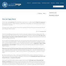
The 10th of the cranial nerves, it is often called the “Nerve of compassion” because when it’s active, it helps create the “warm-fuzzies” that we feel in our chest when we get a hug or are moved by a chick-flick. The vagus nerve is really a bundle of nerves that originates at the top of the spinal cord and enervates all sorts of organs. Here’s a picture: (Peter Jurek, www.peterjurek.com) That first little offshoot stimulates certain muscles in the vocal chamber, facilitating communication.
You can see from this diagram that the vagus nerve branches off to everything from the neck down to the colon. Pretty easy to see how even the simplest yoga routine would stimulate lots of branches of this nerve. But here’s something even cooler – the research that Dacher Ketlner, director of the Social Interaction Laboratory at the University of California, Berkeley is doing shows that stimulating that vagus nerve is not only good for you – it’s good for the planet!
Yoga boosts heart health, new research finds. Heart rate variability, a sign of a healthy heart, has been shown to be higher in yoga practitioners than in non-practitioners, according to research to be published in a forthcoming issue of the International Journal of Medical Engineering and Informatics.

Role Of the Autonomic Nervous System in Emotion. Vagus nerve. Vasovagal response. Signs and symptoms[edit] Episodes of vasovagal response are typically recurrent, and usually occur when the predisposed person is exposed to a specific trigger.
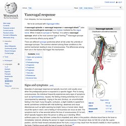
Prior to losing consciousness, the individual frequently experiences early signs of symptoms such as lightheadedness, nausea, the feeling of being extremely hot or cold (accompanied by sweating), ringing in the ears (tinnitus), an uncomfortable feeling in the heart, fuzzy thoughts, confusion, a slight inability to speak/form words (sometimes combined with mild stuttering), weakness and visual disturbances such as lights seeming too bright, fuzzy or tunnel vision, black cloud-like spots in vision, and a feeling of nervousness can occur as well.
The symptoms last for a few seconds before the loss of consciousness (if it is lost), which typically happens when the person is sitting up or standing. The autonomic nervous system's physiologic state (see below) leading to loss of consciousness may persist for several minutes, so: Autonomic nervous system. The autonomic nervous system (ANS or visceral nervous system or involuntary nervous system) is the part of the peripheral nervous system that acts as a control system that functions largely below the level of consciousness to control visceral functions,[1] including heart rate, digestion, respiratory rate, salivation, perspiration, pupillary dilation, micturition (urination), sexual arousal, breathing and swallowing.
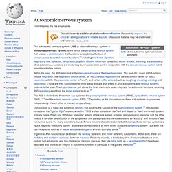
Most autonomous functions are involuntary but they can often work in conjunction with the somatic nervous system which provides voluntary control. Within the brain, the ANS is located in the medulla oblongata in the lower brainstem. The medulla's major ANS functions include respiration (the respiratory control center, or "rcc"), cardiac regulation (the cardiac control center, or "ccc"), vasomotor activity (the vasomotor center or "vmc"), and certain reflex actions (such as coughing, sneezing, vomiting and swallowing).
Anatomy[edit] Sympathetic division[edit] Sympathetic nervous system. The (ortho-) sympathetic nervous system (SNS) is one of three major parts of the autonomic nervous system (the others being the enteric and parasympathetic systems).
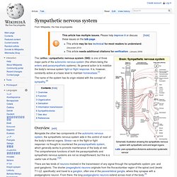
Its general action is to mobilize the body's nervous system fight-or-flight response. It is, however, constantly active at a basic level to maintain homeostasis.[1] The name of the system has its origin related with the concept of sympathy.[2] Overview[edit] There are two kinds of neurons involved in the transmission of any signal through the sympathetic system: pre- and post-ganglionic. At the synapses within the ganglia, preganglionic neurons release acetylcholine, a neurotransmitter that activates nicotinic acetylcholine receptors on postganglionic neurons. Adrenal medulla.
Medullary part of the adrenal gland (on the pointer).
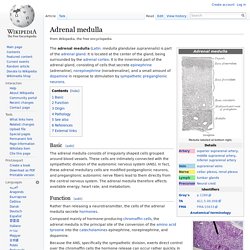
The adrenal medulla (Latin: medulla glandulae suprarenalis) is part of the adrenal gland. It is located at the center of the gland, being surrounded by the adrenal cortex. It is the innermost part of the adrenal gland, consisting of cells that secrete epinephrine (adrenaline), norepinephrine (noradrenaline), and a small amount of dopamine in response to stimulation by sympathetic preganglionic neurons.
Basic[edit] The adrenal medulla consists of irregularly shaped cells grouped around blood vessels. Function[edit] Parasympathetic nervous system. Sympathetic nervous system. Sympathetic nervous system. The sympathetic and the parasympathetic nervous system are parts of what is commonly called the autonomic nervous system.
(Autonomic = can not be controlled by the mind). You can say that these systems work in balance with each other and directly or indirectly affect almost every structure in the body (e.g. heartfrequence, heartcapacity, lumbar function, kidneys, blood vessels, stomach and intestines)