
Motor systems. Motor Pathways All lower motor neuron (LMN) groups except the facial nerve nucleus that supplies the muscles of facial expression consist of both LMNs that supply the skeletal muscle fibers ( ) and LMNs that supply the small contractile elements in the muscle spindles ( ). The muscles of facial expression do not have muscle spindles and are not supplied by gamma LMNs. The alpha LMNs regulate contraction of the skeletal muscles to produce movement. . This mechanism may account for recovery of physiological function in some LMN diseases such as polio. If the alpha-LMN cell body itself is damaged or is in the process of dying (e.g., in amyotrophic lateral sclerosis), the axon may produce aberrant action potentials (agonal bursts of electrical activity) that result in muscle fiber contraction throughout the motor unit, called a fasciculation, which is visually observable. SPINAL TRACTS. DoctorNajeeb. Neuroanatomy Tutorial. Human Physiology - Neurons & the Nervous System II.
BIO 301Human Physiology Neurons & the Nervous System - Part 2 The Human Nervous System consists of the Central Nervous System & the Peripheral Nervous System.

Central Nervous System: 1 - Brain 2 - Spinal cord Peripheral Nervous System: 1 - Cranial nerves (12 pair) & their branches 2 - Spinal nerves (31 pair) & their branches Central Nervous System Evolution of the human brain Source: training.seer.cancer.gov Source: Divisions of Peripheral Nervous System - 1 - Somatic - supplies & receives fibers (neurons) to & from the skin, skeletal muscles, joints, & tendons Used with permission of John Kimball 2 - Visceral - supplies & receives fibers to & from smooth muscle, cardiac muscle, and glands.
Source: NEUROANATOMY I. - Structures of the central nervous system. Interactiv program Authors: Kateřina Kikalová, Libor Machálek, Vladimír Holibka, Miroslav Kutal, Milada Bezděková Program realization: Milan Heřman, Petr Heřman.
M1 Class Page. TIPS (thanks to Feinberg Footnotes) It took several weeks before the material started to make any sense to me at all.

Just be persistent with your studying, and layer the information... Learn the most basic things first, then start to fill in the details. It WILL come together in the end if you work at it. Dr. Behavioral Science—DO NOT MAKE A BIG DEAL OUT OF IT... as long as you read over the lecture notes and the recommended study guide the unit director sends our, you will be fine! Dr. < Back. Posterior view of the brain stem. Neurology « MedicalSchoolResources.com. Medical Neurosciences.
Unit No. 2: Brain stem This site is designed to allow you to view the different pathways of the brain stem in three dimentions. To do this you need to click on the illustration on each page with the words "Play 3D Movie. " You must have Quicktime 5 or greator to view the movies. You can download the latest version of Quicktime from the Apple web site. All material on this website is copyrighted by UW-Madison Medical schooll.
The Cerebellum. PATTS - Nervous System. Don't Be A Salmon: Medicine Archives. Etymology of Neuroscience Terms. THE BRAIN FROM TOP TO BOTTOM. The basic function of the brain is to produce behaviours, which are, first and foremost, movements.
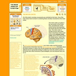
Several different regions of the cerebral cortex are involved in controlling the body's movements. These regions are organized into a hierarchy like the crew of a ship. On an ancient galley, for example, the captain determined the destination for a voyage by assessing the various factors that might make such a trip worthwhile. Then his lieutenants calculated the direction that the ship had to travel to reach that destination, based on weather conditions. End-plate potential. Endplate potential (EPP) and mEPPs recorded from a frog muscle fiber End plate potentials (EPPs) are the depolarizations of skeletal muscle fibers caused by neurotransmitters binding to the postsynaptic membrane in the neuromuscular junction.
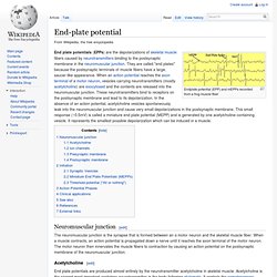
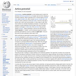
They are called "end plates" because the postsynaptic terminals of muscle fibers have a large, saucer-like appearance. Action potential. As an action potential travels down the axon, there is a change in polarity across the membrane.
Receptor potential. Receptor potential, a type of graded potential, is the transmembrane potential difference of a sensory receptor.[1] An example of a receptor potential is in a taste bud, where taste is converted into an electrical signal sent to the brain.

When stimulated, the taste bud triggers the release of neurotransmitter through exocytosis of synaptic vesicles from the presynaptic membrane. The neurotransmitter molecules diffuse across the synaptic cleft to the postsynaptic membrane of the primary sensory neuron, where they elicit an action potential. See also[edit] References[edit] Jump up ^ Hille, Bertil (2001). Synaptic potential. Synaptic potential refers to the difference in voltage between the inside and outside of a postsynaptic neuron.
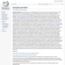
In other words, they are the “incoming” signal of a neuron. Synaptic potential comes in two forms: excitatory and inhibitory. Neurons, Synapses, Action Potentials, and Neurotransmission - The Mind Project. Function of neurons Because our main interest lies in exploring how information processing occurs in the brain, we are going to ignore glia.
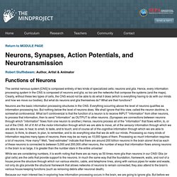
But before we see how neurons process information (and what that means), you need to know a few things about the structure of neurons. Structure of neurons While there are as many as 10,000 specific types of neurons in the human brain, generally speaking, there are three kinds of neurons: motor neurons (for conveying motor information), sensory neurons (for conveying sensory information), and interneurons (which convey information between different types of neurons). The following image identifies how neurons come in various shapes and sizes. Neuronal signaling Conduction Neurotransmission. Chemical synapse. Illustration of the major elements in chemical synaptic transmission.
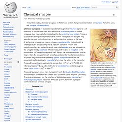
An electrochemical wave called an action potential travels along the axon of a neuron. When the action potential reaches the presynaptic terminal, it provokes the release of a small quantity of neurotransmitter molecules, which bind to chemical receptor molecules located in the membrane of another neuron, the postsynaptic neuron, on the opposite side of the synaptic cleft. Chemical synapses are specialized junctions through which neurons signal to each other and to non-neuronal cells such as those in muscles or glands. Chemical synapses allow neurons to form circuits within the central nervous system. They are crucial to the biological computations that underlie perception and thought. The adult human brain is estimated to contain from 1014 to 5 × 10¹⁴ (100–500 trillion) synapses.[1] Every cubic millimeter of cerebral cortex contains roughly a billion (short scale, i.e. 109) of them.[2] Structure[edit] Overview[edit]