
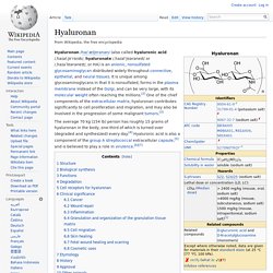
Regeneration: what does it mean and how does it work? Glial fibrillary acidic protein. Vimentin. Immunofluorescence staining pattern of vimentin antibodies.
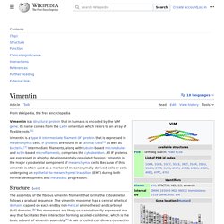
Produced by incubating vimentin primary antibodies and FITC labelled secondary antibodies with HEp-20-10 cells. Vimentin is a protein that in humans is encoded by the VIM gene. Testing selected optimal descriptors with artificial neural networks - RSC Advances. If you are not the author of this article and you wish to reproduce material from it in a third party non-RSC publication you must formally request permission using RightsLink.
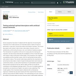
Go to our Instructions for using RightsLink page for details. Authors contributing to RSC publications (journal articles, books or book chapters) do not need to formally request permission to reproduce material contained in this article provided that the correct acknowledgement is given with the reproduced material. Reproduced material should be attributed as follows: For reproduction of material from NJC: Reproduced from Ref.
XX with permission from the Centre National de la Recherche Scientifique (CNRS) and The Royal Society of Chemistry. UConn unveils ambitious knee regeneration research. The University of Connecticut announced the launch of its new grand research challenge: regeneration of a human knee within 7 years, and an entire limb within 15 years.
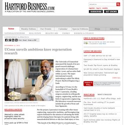
The major international research undertaking is called The HEAL Project: Hartford Engineering a Limb. According to UConn, it is the brainchild of UConn Health's Cato T. Laurencin, a leading surgeon-scientist in orthopaedic surgery, engineering, and the new field of regenerative engineering. Regeneration: what does it mean and how does it work? Regeneration and humans Regeneration means the regrowth of a damaged or missing organ part from the remaining tissue.
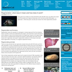
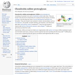
As adults, humans can regenerate some organs, such as the liver. If part of the liver is lost by disease or injury, the liver grows back to its original size, though not its original shape. Hyaluronan. Hyaluronan /haɪˈæljʊrənən/ (also called hyaluronic acid /ˌhaɪəl.jʊˈrɒnɨk/, hyaluronate /ˌhaɪəlˈjʊərəneɪt/ or /ˌhaɪəˈlʊərəneɪt/, or HA) is an anionic, nonsulfated glycosaminoglycan distributed widely throughout connective, epithelial, and neural tissues.
It is unique among glycosaminoglycans in that it is nonsulfated, forms in the plasma membrane instead of the Golgi, and can be very large, with its molecular weight often reaching the millions.[2] One of the chief components of the extracellular matrix, hyaluronan contributes significantly to cell proliferation and migration, and may also be involved in the progression of some malignant tumors.[3] Structure[edit]
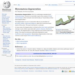
Chondroitin sulfate proteoglycan. Structure of the protein core of aggrecan or chondroitin sulfate proteoglycan 1 Chondroitin sulfate proteoglycans (CSPGs) are proteoglycans consisting of a protein core and a chondroitin sulfate side chain.
They are known to be structural components of a variety of human tissues, including cartilage, and also play key roles in neural development and glial scar formation. They are known to be involved in certain cell processes, such as cell adhesion, cell growth, receptor binding, cell migration, and interaction with other extracellular matrix constituents.[1] They are also known to interact with laminin, fibronectin, tenascin, and collagen.[1] CSPGs are generally secreted from cells.
Importantly, CSPGs are known to inhibit axon regeneration after spinal cord injury. CSPGs contribute to glial scar formation post injury, acting as a barrier against new axons growing into the injury site.[2] CSPGs play a crucial role in explaining why the spinal cord doesn't self-regenerate after an injury.
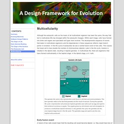
Myxomatous degeneration. Myxomatous degeneration refers to a pathological weakening of connective tissue.
The term is most often used in the context of mitral valve prolapse, which is known more technically as "myxomatous mitral valve degeneration. " The degeneration occurs in conjunction with an accumulation of dermatan sulfate, a glycosaminoglycan, within the connective tissue matrix of the valve. The exact mechanism is unknown. Multicellularity - A Design Framework for Evolution. Although the eukaryotic cells as the masis of all multicellular organism has been the same, the way that led to multicellula rlife has diverged within the eukaryotic lineages.
Within each linage, cells have formed into limbs and organs and specialied cell types have evolved. The developmental sequence of events that leads to multicellular organisms and the dependecies in these sequences reflects major branch points in evolution. In the life cycle of eukaryotes we see a central fusion event of two cells. This causes the fused cell to have double the number of chromosomes (diploid). Phosphodiesterase. A phosphodiesterase (PDE) is any enzyme that breaks a phosphodiester bond.

Usually, people speaking of phosphodiesterase are referring to cyclic nucleotide phosphodiesterases, which have great clinical significance and are described below. However, there are many other families of phosphodiesterases, including phospholipases C and D, autotaxin, sphingomyelin phosphodiesterase, DNases, RNases, and restriction endonucleases (which all break the phosphodiester backbone of DNA or RNA), as well as numerous less-well-characterized small-molecule phosphodiesterases. The cyclic nucleotide phosphodiesterases comprise a group of enzymes that degrade the phosphodiester bond in the second messenger molecules cAMP and cGMP. Transducin. Sensory rhodopsin II (rainbow colored) embedded in a lipid bilayer (heads red and tails blue) with Transducin below it.
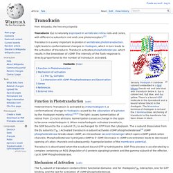
Gtα is colored red, Gtβ blue, and Gtγ yellow. There is a bound GDP molecule in the Gtα-subunit and a bound retinal (black) in the rhodopsin. The N-terminus terminus of rhodopsin is red and the C-terminus blue. AccessSurgery - Print. Surgical Anatomy Knowledge of detailed cardiac anatomy is a prerequisite for successful surgery. Nowhere is this more important than in the setting of congenital cardiac malformations. —R.H. Anderson, B.R. AccessSurgery - Print. Search: Periostin. Untitled. My research interests focus on cellular and molecular mechanisms underlying early cardiac cell specification, differentiation, and heart organogenesis. Specifically, we target the cardiac precursor cell population. Currently, we are analyzing effects of alcohol inducing Fetal Alcohol Syndrome, of lithium and homocysteine, and the mechanism by which folate and with even greater efficacy, folate/myo-inositol combination prevents cardiac birth defects induced by these environmental factors.
These studies have been extended into aspects of wound healing.
Untitled. Katyaxolotl. Objective This experiment will seek to demonstrate regulative development in Ambystoma mexicanum embryos. Specifically, we will split the morphogenetic field that is responsible for heart formation using grafted tissue from the gill area, and attempt to form two hearts – one on either side of the grafted tissue. Background. TGF beta signaling pathway. The transforming growth factor beta (TGFB) signaling pathway is involved in many cellular processes in both the adult organism and the developing embryo including cell growth, cell differentiation, apoptosis, cellular homeostasis and other cellular functions. In spite of the wide range of cellular processes that the TGFβ signaling pathway regulates, the process is relatively simple. TGFβ superfamily ligands bind to a type II receptor, which recruits and phosphorylates a type I receptor.
The type I receptor then phosphorylates receptor-regulated SMADs (R-SMADs) which can now bind the coSMAD SMAD4. R-SMAD/coSMAD complexes accumulate in the nucleus where they act as transcription factors and participate in the regulation of target gene expression. Mechanism[edit] Ligand binding[edit] Randal Voss - Ambystoma Genetic Stock Center at UK. Axolotl Embryo Staging Series. S - Embryo Photo Log. Ambystoma Genetic Stock Center. Welcome to Ambystoma Genetic Stock Center. Book - The Frog Its Reproduction and Development 11 - Embryology. Cardiac Embryology - Embryology. How Salamanders Sprout New Limbs. PLOS ONE: Transcriptomic Analysis of Endangered Chinese Salamander: Identification of Immune, Sex and Reproduction-Related Genes and Genetic Markers.
Periostin. Fasciclin domain. In molecular biology, the fasciclin domain (FAS1 domain) is an extracellular domain of about 140 amino acid residues. Fasciclin 2. Matricellular protein. Genome size, cell size, and the evolution of enucleated erythrocytes in attenuate salamanders. Salamander Biology – Anatomy and Physiology. Hedgehog Signaling Controls Dorsal Ventral Patterning and Induction of Axolotl Tail Regeneration. S - Rearing from Egg to Adult.
Regulative Development in Axolotl Embryos; Splitting the Heart Field - 发育生物学 - 资讯 - 生物在线. Cardiovascular System Development - Embryology. S - Embryo Photo Log. Developmental Biology Interactive. Fibroblasts are the Critical Cells in Salamander Limb Regeneration » UC Irvine Limb Regeneration. McGowan Institute for Regenerative Medicine. Fossil has evidence of limb regeneration in 300 million year old amphibian. Making News » UC Irvine Limb Regeneration. Mexican Axolotl Provides Insights into Potential of Human Regenerative Medicine. Regeneration: what does it mean and how does it work? S - Requirements & Water Conditions in Captivity. ZFIN Figure: Mugoni et al., 2013, Fig. 1. Caudata Culture Species Entry - Crested Triturus species.