
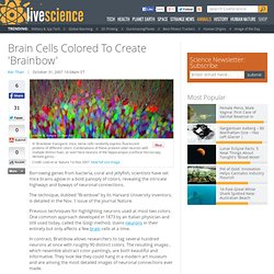
Fragment of rat brain simulated in supercomputer. A simulated brain slice from the Blue Brain Project: neurons are coloured according to their levels of electrical activity.
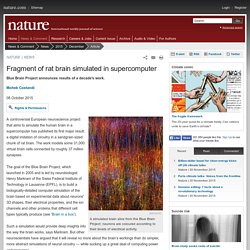
A controversial European neuroscience project that aims to simulate the human brain in a supercomputer has published its first major result: a digital imitation of circuitry in a sandgrain-sized chunk of rat brain. The work models some 31,000 virtual brain cells connected by roughly 37 million synapses. The goal of the Blue Brain Project, which launched in 2005 and is led by neurobiologist Henry Markram of the Swiss Federal Institute of Technology in Lausanne (EPFL), is to build a biologically-detailed computer simulation of the brain based on experimental data about neurons' 3D shapes, their electrical properties, and the ion channels and other proteins that different cell types typically produce (see ‘Brain in a box’).
Such a simulation would provide deep insights into the way the brain works, says Markram. But Markram is undeterred. New Brain Scans Show How Dolphins Use Sound to See. ?articles. Dissecting how the brain works is tricky.
Genetic engineering techniques allow researchers to tag neurons of interest with fluorescent markers that glow upon neural activation, but capturing those brain areas in action, both as distinct neural circuits and at the resolution of single cells, can be hard. Studying the neural activity of Drosophila, for example, involves microsurgery to remove the top of a fly’s head to get a clear view of its brain, a task so delicate that only practiced technicians with steady hands are able to complete it successfully. To monitor brain activity in mice and other animals, neuroscientists often rely on a well-established technique called patch clamping, which can ignite career-questioning frustration, as electrical noise spoils seemingly good data and cells begin to die after only a few seconds of recording.
Brain Simulation. Fast whole-brain imaging made possible for the first time with LSFM - volume render. Untitled. Brain function relies on communication between large populations of neurons across multiple brain areas, a full understanding of which would require knowledge of the time-varying activity of all neurons in the central nervous system.

Here we use light-sheet microscopy to record activity, reported through the genetically encoded calcium indicator GCaMP5G, from the entire volume of the brain of the larval zebrafish in vivo at 0.8 Hz, capturing more than 80% of all neurons at single-cell resolution. Demonstrating how this technique can be used to reveal functionally defined circuits across the brain, we identify two populations of neurons with correlated activity patterns. One circuit consists of hindbrain neurons functionally coupled to spinal cord neuropil. Subject terms: Computational neuroscience• Light-sheet microscopy Figures Videos.
Watch chemicals turn into memories - the first time this has ever been recorded. Scientists have known for a while that our memories are the process are synaptic transmissions in our brain and are stored in neurons, but they have been able to film the actual process for the first time inside of a mouse.
This groundbreaking video was made in the lab of Robert Singer of Albert Einstein College of Medicine and the paper describing the process was published in Science. Our lives are defined by our memories. Everything we do, including driving a car, laughing at jokes, cooking dinner, or calling a friend relies on our prior knowledge of similar events. But how is all of this created and stored in our brains? Memory research is extremely complex, but neuroscientists have been able to work a lot of it out. Memories are made by messenger RNA (mRNA) that encode β-actin protein. Because the process is so delicate, it has been incredibly difficult for researchers to see it happen in real time.
This computer took 40 minutes to simulate one second of brain activity. Scientists can now turn brains invisible. Proteins don't need to be in their native conformation to study them using antibody based imaging techniques most of the time, however, for those antibodies that were raised against whole protein rather than an exposed epitope tissues like this can be treated with citrate and heat (or other antigen recovery techniques) to restore epitope binding activity.
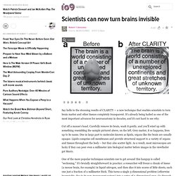
CLARITY should be compatible with antigen recovery. Other than this, proteins don't really need to be in their active conformations for this kind of study, however, the researchers that developed this technique tested it with brains expressing GFP (and I believe, either Tomato or mCherry) which retained fluorescence after treatment. This is also true of standard PFA fixed tissue, fixation protocols are fairly carefully calibrated to prevent "over-fixing" which does adversely affect this kind of thing. Although I didn't work on CLARITY its self, I do work in this field. and if you have more questions I'd be happy to answer them. Brain Cells Colored To Create 'Brainbow' Borrowing genes from bacteria, coral and jellyfish, scientists have set mice brains aglow in a bold panoply of colors, revealing the intricate highways and byways of neuronal connections.
The technique, dubbed "Brainbow" by its Harvard University inventors, is detailed in the Nov. 1 issue of the journal Nature. Previous techniques for highlighting neurons used at most two colors. Babies' brains to be mapped in the womb and after birth. 9 April 2013Last updated at 21:45 ET By Smitha Mundasad Health reporter, BBC News Scientists are drawing up a map of the growing baby's brain UK scientists have embarked on a six-year project to map how nerve connections develop in babies' brains while still in the womb and after birth.
By the time a baby takes its first breath many of the key pathways between nerves have already been made. And some of these will help determine how a baby thinks or sees the world, and may have a role to play in the development of conditions such as autism, scientists say. Major Brain Dump! Human Connectome. Human Brain Project - Home. BigBrain. Reverse Engineering The Brain. This is part of IEEE Spectrum's SPECIAL REPORT: THE SINGULARITY PHOTO: Timothy Archibald What do fruit-fly brains have in common with microchips?
That's not the setup for a bad joke; it's David Adler's life. Under Adler's ultra sophisticated electron beam microscopes, advanced microprocessors with transistors far smaller than red blood cells have been reduced to their wiring diagrams. Mapping the human brain connectivity. 70,000+ Have Played ‘Eyewire’ Game That Trains Computers To Map the Brain. Your connectome, the map of all 86 billion connected neurons in your brain, is hopelessly complex.

In fact, one human connectome has a staggering 10,000 times that number of neural pathways. Every thought you have and every memory you hold exists in your connectome, and major efforts are under way to map it. The good news is that you don’t need a fancy neuroscience degree to help out. In fact, the fancy degreed neuroscientists are hoping that you might pitch in. Created by scientists at MIT, Eyewire is a browser game that lets players take on the challenge of mapping neural pathways in brains — no scientific background required.
In an amplifying way, the team at MIT hopes that these human assisted computers will one day learn to map neurons by themselves. To date, over 70,000 gamers from over 100 countries have signed up to play Eyewire, and it’s a good thing they did. Mapping the connectome will be considerably more challenging than mapping the genome, but the lesson learned holds true. Europe's $1.6 Billion Human Brain Project Is On The Verge Of Collapse. Expand The highly ambitious Human Brain Project — an effort to simulate the human brain in a supercomputer — is drawing fire from neuroscientists who say the initiative is unfeasible, unfocused, and unaccountable.
Europe's Human Brain Project was launched early last year — an effort to develop better tools to study how the brain works. As part of the initiative, researchers will take the next ten years to understand and map the network of over a hundred billion neuronal connections that elicit emotions, volitional thought, and even consciousness itself.
And to do so, the researchers are planning to use a progressively scaled-up multilayered simulation running on a supercomputer. More than 80 European and international research institutions are taking part. But now, hundreds of neuroscientists (yes, hundreds — the crisis is that bad) have signed an open letter condemning what they perceive as an absence of feasibility and transparency. Are Efforts at Total Brain Simulation Putting the Cart Before the Horse? Since it was awarded a one billion euro, decade-long research grant last year, the Human Brain Project has been the center of extreme excitement and heavy criticism. The project aims to simulate the human brain in silicon on a yet-to-be-assembled supercomputer of massive computational power.
The goal? Understanding. In a recent paper (more below), HBP researchers write, “The ultimate prime aim is to imitate and understand the native computations, algorithms, states, actions, and emergent behavior of the brain, as well as promote brain-inspired technology.” A Worm's Mind In A Lego Body. Take the connectome of a worm and transplant it as software in a Lego Mindstorms EV3 robot - what happens next? It is a deep and long standing philosophical question.
Are we just the sum of our neural networks. Of course, if you work in AI you take the answer mostly for granted, but until someone builds a human brain and switches it on we really don't have a concrete example of the principle in action. KDS444, modified by Nnemo The nematode worm Caenorhabditis elegans (C. elegans) is tiny and only has 302 neurons.
The model is accurate in its connections and makes use of UDP packets to fire neurons. The software works with sensors and effectors provided by a simple LEGO robot.