
Marian Koshland Science Museum. 3D Brain. Copyright © Society for Neuroscience (2017).
Users may copy images and text, but must provide attribution to the Society for Neuroscience if an image and/or text is transmitted to another party, or if an image and/or text is used or cited in User’s work. Brain: Atlas of human anatomy with MRI. The module on the anatomy of the brain based on MRI with axial slices was redesigned, having received multiple requests from users for coronal and sagittal slices.
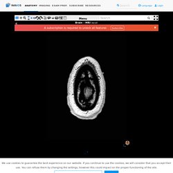
The elaboration of this new module, its labeling of more than 524 structures on 379 MRI images in three different views and on 26 anatomical diagrams, took more than 6 months. This module is intended for all physicians and non-physicians with an interest in neuroanatomy and medical imaging, particularly for general practitioners and specialists in neurology, neurosurgery, anatomy and neurosciences, as well as speech pathologists and psychomotor therapists. Anatomy of the encephalon in MRI (axial, coronal and sagittal slices) Cerebral images used for this module on human anatomy An MRI was performed in thin slices (0.6 mm) on a healthy individual, with volumetric 3D imaging using T1 weighting without injection of gadolinium in the three normally used views, with a matrix of 320/320 pixels, using an MRI machine of 1.5 Tesla.
3-D Brain App - CSHL DNA Learning Center. Apps for iPhone, iPad, and Android devices The 3D Brain is also available as an app for iPhone/iPod/iPad, Android, and Windows Phone devices!
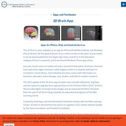
All the great features of our online version in the palm of your hand! It can be downloaded from the Apple App Store; look for it in the Education category (iTunes is required), and Android and Windows Phone app stores. Use your touch screen to rotate and zoom around 29 interactive structures. Anatomy of the Human Brain 2019 - 3D model by INTERVOKE (@intervoke) [4870387] Researchers uncover drain pipes in our brains. Digital Anatomist Interactive Atlases.
Structural Informatics GroupDepartment of Biological StructureUniversity of Washington Seattle, Washington, USA Atlases Content: 2-D and 3-D views of the brain from cadaver sections, MRI scans, and computer reconstructions.Author: John W.
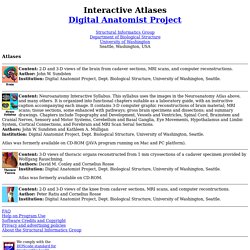
SundstenInstitution: Digital Anatomist Project, Dept. Biological Structure, University of Washington, Seattle. Content: Neuroanatomy Interactive Syllabus. Atlas was formerly available on CD-ROM (JAVA program running on Mac and PC platform). Content: 3-D views of thoracic organs reconstructed from 1 mm cryosections of a cadaver specimen provided by Wolfgang Rauschning.Authors: David M. Atlas was formerly available on CD-ROM. Neuroanatomy Tutorial. Neuroanatomy—A Primer. The human brain is a unique structure that boasts a complex three-dimensional architecture.
Neuroscientists are only beginning to understand how the different parts of this intricate configuration work together to produce behavior. In the numerous neuroimaging studies that are published weekly, researchers use common neuroanatomical terms to denote location, organization, and, at times, implied function. Though a complete discussion of neuroanatomy is worthy of a thick textbook full of elaborate illustrations, common terminology used in neuroscientific research is highlighted below.
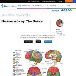
The basics Perched on top of the spinal column, the brain is the epicenter of the human nervous system. The cerebral cortex is divided into two hemispheres connected by the corpus callosum, a bridge of wide, flat neural fibers that act as communication relays between the two sides. Credit: Nucleus Medical Art, Inc. Click image for larger view. Neuroanatomy Learning Modules. Allen Brain Atlas: Human Brain.
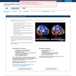
Windows Minimum Configuration Operating System: Microsoft Windows 7CPU: Intel Core Duo or AMD 1.8GHzSystem Memory: 1GBGraphics Card: Hardware 3D OpenGL accelerated AGP or PCI Express with 64MB RAMScreen: 1024x768, 32-bit true colorHard Disk: 200MB free space Note: The Brain Explorer 2 software is known to work with the following video chipsets: nVidia GeForce 9400/9600, nVidia Quadro FX 1800/3800/5600, AMD Radeon 9600, AMD Radeon HD 3200/4550, Intel Q35/Q45 Express Important: Please install the latest drivers for your video card for best compatibility and performance.
3D Brain. Brain Function, Anatomy & Diagram. Much of the brain's job involves receiving information from the rest of the body, interpreting that information, and then guiding the body's response to it.
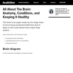
Types of input the brain interprets include odors, light, sounds, and pain. The brain also helps perform vital operations such as breathing, maintaining blood pressure, and releasing hormones. The brain is divided into sections. These sections include the cerebrum, the cerebellum, the diencephalon, and the brain stem. Brain: Brodmann Areas. Brodmann Areas (BA), 10-20 Sites, Primary Functions ADD Centre, Biofeedback Institute of Toronto, American Applied NeuroScience Institute Michael Thompson, MD, James Thompson, PhD., Wu Wenqing, MD.
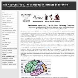
What follows is a project-in-process. It is being done to assist the staff at our centers in their work using EEG Biofeedback (neurofeedback). It is to be a part of The Companion to The Neurofeedback Book which is being written at this time. If it can be a helpful starting point for others, then they are welcome to use it. Each BA has many different functions associated with it. Functions for each Brodmann area are outlined below. NOTE: Please do not get confused. Interactive Brain. BrainInfo. Allen Brain Atlas: Human Brain. Pocket Brain Version 1.0 – Review By Areo Saffarzadeh, MS3.
This review is intended to provide an overview of Pocket Brain, the new interactive neuroanatomy app by eMedia and compare its features to other other iOS based neuroanatomy apps.

In short, Pocket Brain is the most comprehensive, interactive, and functional neuroanatomy app out there. It is worth the investment of any student studying neuroanatomy. Neuroanatomy as a Class To be good at neuroanatomy a student needs decent visual-spatial reasoning skills. Some are gifted in this regard others are not. Brain Geography. The Brain Observatory. The Whole Brain Atlas.
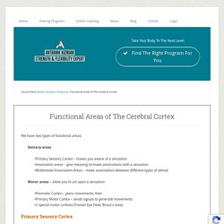
CH 12 Functional Areas of the Cerebral Cortex. Functional Areas of The Cerebral Cortex. We have two types of functional areas: Sensory areas •Primary Sensory Cortex – makes you aware of a sensation •Association areas – give meaning to/make associations with a sensation •Multimodal Association Areas – make associations between different types of stimuli Motor areas – allow you to act upon a sensation •Premotor Cortex – plans movements; then •Primary Motor Cortex – sends signals to generate movements •2 special motor cortices (Frontal Eye Field, Broca’s area) Primary Sensory Cortex For each of the major senses, there is an area called the primary sensory cortex.
Anatomy and Function. 3D Slicer. Structure: precentral gyrus. Chapter 1: Introduction. An understanding of functional neuroanatomy is critical to understanding the symptoms of nervous system damage. Most disorders of the nervous system either target particular brain structures or target components of functional systems. Therefore, knowing these structures and their basic functions permits localization of the nervous system damage. This chapter will consider the important elements of clinical neuroanatomy. There are several good texts that provide greater detail on these systems (1-3). An atlas of brain structures is essential to the study of neuroanatomy. This review will discuss the cellular components of the nervous system first and then will consider the peripheral nervous system (PNS) before discussing the Central nervous system (CNS). 1. 2. 3. 4.
Chapter 9: Limbic System. The limbic system is a convenient way of describing several functionally and anatomically interconnected nuclei and cortical structures that are located in the telencephalon and diencephalon. These nuclei serve several functions, however most have to do with control of functions necessary for self preservation and species preservation. They regulate autonomic and endocrine function, particularly in response to emotional stimuli. They set the level of arousal and are involved in motivation and reinforcing behaviors. Chapter 11: The Cerebral Cortex. General Organization The cerebral cortex is the outer covering of gray matter over the hemispheres.
This is typically 2- 3 mm thick, covering the gyri and sulci. Certain cortical regions have somewhat simpler functions, termed the primary cortices. These include areas directly receiving sensory input (vision, hearing, somatic sensation) or directly involved in production of limb or eye movements. The association cortices subserve more complex functions. Cortex - cortex.pdf. Focus on Brain Disorders. Allen Institute for Brain Science: Home. Interactive Tour of the Brain. Scalable Brain Atlas - Neuroanatomy at your fingertips.
Digital Anatomist Interactive Atlases. BrainNavigator: Elsevier's 3D brain anatomy tool that helps scientists further understand the human brain. The Secret Life of the Brain : 3-D Brain Anatomy. Brain Atlas - 3D Brain Model of Anatomy & Injuries. Traumatic Brain Injury Rehabilitation Educational Resources > FINR Educational Materials > FINR Brain Atlas - 3D Brain Model 3D Brain Model - Explore our interactive 3-dimensional brain atlas to discover where structures are located within the brain, their purpose, and explore brain injury models. All structures and models are accompanied by easy-to-understand detailed explanations. It is our hope that this model assists to further the understanding of brain injury by those affected. Find the FINR 3D Brain model application for the iPhone, iPod Touch, and the iPad in the iTunes App Store.
Click here to launch the FINR Atlas of Brain Injury & Anatomy. FINR Brain Atlas App is Awarded the 18th Annual Communicator - Silver Award of Distinction in the Health & Wellness Mobile Apps Category. 3d Reconstruction of a brain from MRI data. New Account Request. Nervous System Intro. Humans, like all living organisms, can respond to their environment. Humans have two complimentary control systems to do this: the nervous system and the endocrine (hormonal) system. The human nervous system controls everything from breathing and producing digestive enzymes, to memory and intelligence.
Nerve Cells [back to top] UBC Undergraduate Medicine: Neuroanatomy. BRAIN-MAPS-API - BRAINMAPS.ORG - BRAIN ATLAS, BRAIN MAPS, BRAIN STRUCTURE, NEUROINFORMATICS, BRAIN, STEREOTAXIC ATLAS, NEUROSCIENCE. The Brain Maps API is a lightweight multiresolution image viewer with customizable label overlays that lets you embed BrainMaps images in your own web pages with JavaScript, and that can also be used with your own multiresolution images. Compare Brains. Comparative Mammalian Brain Collections. Why Comparative Neurobiology? Why have I chosen to create a website entirely dedicated to the neuroanatomy and related issues of a number of different species?
Why not focus just on humans? What is the value in comparison studies? Mouse Brain Mapped in Greatest Detail Yet. 10 Great Sites for Reviewing Brain Anatomy. I’ve been absolutely immersed in brain anatomy (which I now heart) for the past eight months. In the process I’ve amassed a rather large collection of links. Kimball's Biology Pages. Ways to Search These Pages Search Engine.
Enter desired term(s) in box above right and click on "GO".