
Diagnostic Value of Breast Proton Magnetic Resonance Spectroscopy at 1.5T in Different Histopathological Types. The purpose of this study was to investigate the usefulness of quantitative proton magnetic resonance spectroscopy (1H-MRS) for characterizing breast lesions at 1.5T, and to evaluate the diagnostic performance of in vivo breast 1H-MRS using receiver operating characteristics (ROC) analysis. 112 patients (99 malignant and 13 benign tumors) who were scanned with the MRI/MRS protocol were included in this study.
Choline-containing compounds (tCho) levels were measured and compared with histological findings. The measured tCho levels in this work had range of 0.08–9.99 mmol/kg from 65 (66%) of 99 patients with malignant tumors. Of the 13 benign lesions, 1H-MRS detected one as false positive, with tCho level of 0.66 mmol/kg. The optimal tCho level cutoff point that yielded the highest accuracy was found to be >0.0 mmol/kg. The resulting sensitivity was 66% and specificity 92% for distinguishing benign from malignant lesions. Diagnosing inborn errors of lipid metabolism with proton nuclear magnetic resonance spectroscopy. - Free Online Library. Proton nuclear magnetic resonance ([sup.1]H-NMR) [3] spectroscopy is a versatile technique that can be used in a wide range of disciplines.
The best known medical applications are in vivo magnetic resonance imaging and spectroscopy, but in vitro [sup.1]H-NMR spectroscopy of body fluids has also been used to diagnose inborn errors of metabolism (1-3). In contrast to conventional techniques, NMR can detect the majority of all proton-containing metabolites in a single experiment lasting -15 min. Furthermore, it is a nondestructive method and requires little or no sample preparation. Neurometabolic alterations in bipolar disorder with anxiety symptoms: A proton magnetic resonance spectroscopy study of the prefrontal whiter matter. Ahajournals. Psychiatry Online. Proton magnetic resonance spectroscopy assessment of metabolite status. Takahiro Ito,1 Sachiko Tanaka-Mizuno,2,3 Narihito Iwashita,4 Ikuo Tooyama,5 Akihiko Shiino,6 Katsuyuki Miura,1,7 Sei Fukui4 1Department of Public Health, Shiga University of Medical Science, 2Department of Medical Statistics, Shiga University of Medical Science, Otsu, Japan; 3The Center for Data Science Education and Research, Shiga University, Hikone, Japan; 4Department of Anesthesiology, Interdisciplinary Pain Management Center, Shiga University of Medical Science Hospital, 5Molecular Neuroscience Research Center, Shiga University of Medical Science, 6Biomedical MR Science Center, Shiga University of Medical Science, 7Center for Epidemiologic Research in Asia, Shiga University of Medical Science, Otsu, Japan Background: Chronic pain is a common cause of reduced quality of life.

Recent studies suggest that chronic pain patients have a different brain neurometabolic status to healthy people. Introduction Materials and methods. - Brasil - Proton magnetic resonance spectroscopy of the frontal lobe in schizophrenics: a critical review of the methodology Proton magnetic resonance spectroscopy of the frontal lobe in schizophrenics: a critical review of the methodology. Proton magnetic resonance spectroscopy of the frontal lobe in schizophrenics: a critical review of the methodology Espectroscopia de próton por ressonância magnética de lobo frontal em esquizofrênicos revisão crítica da metodologia Rafael Faria Sanches; José Alexandre de Souza Crippa; Jaime Eduardo Cecílio Hallak; David Araújo; Antonio Waldo Zuardi From the Departments of Neuropsychiatry and Medical Psychology, Faculty of Medicine of Ribeirão Preto, University of São Paulo - São Paulo/SP, Brazil.
E-mail: rfsanches@uol.com.br Schizophrenic patients undergoing proton magnetic resonance spectroscopy show alterations in N-acetyl aspartate levels in several brain regions, indicating neuronal dysfunction. The neurochemical basis of human cortical auditory processing: combining proton magnetic resonance spectroscopy and magnetoencephalography. 1.Fruhstorfer H, Soveri P, Järvilehto T: Short-term habituation of the auditory evoked response in man.
Electroencephalogr Clin Neurophysiol. 1970, 28 (2): 153-161. 10.1016/0013-4694(70)90183-5.Article CAS PubMed Google Scholar 2.Hari R, Kaila K, Katila T, Tuomisto T, Varpula T: Interstimulus interval dependence of the auditory vertex response and its magnetic counterpart: implications for their neural generation. Electroencephalogr Clin Neurophysiol. 1982, 54 (5): 561-569. 10.1016/0013-4694(82)90041-4.Article CAS PubMed Google Scholar 3.Okamoto H, Kakigi R, Gunji A, Kubo T, Pantev C: The dependence of the auditory evoked N1m decrement on the bandwidth of preceding notch-filtered noise.
Eur J Neurosci. 2005, 21 (7): 1957-1961.Article CAS PubMed Google Scholar 4.Michael N, Ostermann J, Sörös P, Schwindt W, Pfleiderer B: Altered habituation in the auditory cortex in a subgroup of depressed patients by functional magnetic resonance imaging. The role of proton magnetic resonance spectroscopy in the diagnosis and categorization of cerebral abscesses in: Neurosurgical Focus Volume 24 Issue 6 (2008) Journals.asm. Hub of NMR, IR, Mass and UV-Visible Spectroscopy.
Chemical compound - Proton magnetic resonance spectroscopy. Proton NMR spectra yield a great deal of information about molecular structure because most organic molecules contain many hydrogen atoms, and the hydrogen atoms absorb energy of different wavelengths depending on their bonding environment.

NMR absorbances appear in a spectrum as a series of sharp spikes or peaks. Although there is no vertical scale on the spectrum, the relative height of each peak corresponds roughly to the strength of the absorption. The horizontal scale does not show proton resonances in simple wavelength units. Instead, the position of each peak is normally measured relative to the absorption of the protons in the compound tetramethylsilane, (CH3)4Si. Introduction to proton NMR. Proton Nuclear Magnetic Resonance Spectroscopy. Proton NMR Spectroscopy - Some basic point discussion Over the past fifty years nuclear magnetic resonance spectroscopy, commonly referred to as nmr, has become the preeminent technique for determining the structure of organic compounds.

Of all the spectroscopic methods, it is the only one for which a complete analysis and interpretation of the entire spectrum is normally expected. Although larger amounts of sample are needed than for mass spectroscopy, nmr is non-destructive, and with modern instruments good data may be obtained from samples weighing less than a milligram. Radiology Reference Article. The technique of magnetic resonance spectroscopy (usually shortened to MR spectroscopy or MRS) allows tissue to be interrogated for the presence and concentration of various metabolites.
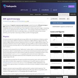
Grossman and Yousem said "If you need this to help you, go back to page 1; everything except Canavan (disease) has low NAA, high choline" 1. This is perhaps a little harsh, however, it is fair to say that MRS often does not add a great deal to an overall MR study but does increase specificity, and may help in improving our ability to predict histological grade. The basic principle that enables MR spectroscopy (MRS) is that the distribution of electrons within an atom cause nuclei in different molecules to experience a slightly different magnetic field.
This results in slightly different resonant frequencies, which in turn return a slightly different signal. Proton nuclear magnetic resonance spectroscopy of aqueous solutions: complete elimination of the water resonance by spin-spin relaxation. Pay to View. Proton magnetic resonance spectroscopy changes after lithium treatment. Systematic review. Abstract 1H MRS is widely used in the research of mental disorders.
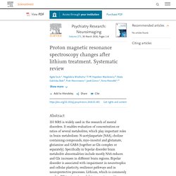
It enables evaluation of concentration or ratios of several metabolites, which play important roles in brain metabolism: N-acetylaspartate (NAA), choline containing compounds, myo-inositol and glutamate, glutamine and GABA (together as Glx complex or separately). Pubs.rsna. Moving the goal posts: enhancing the sensitivity of PARASHIFT proton magnetic resonance imaging and spectroscopy - Chemical Science. Mobility of Water in Wheat Flour Suspensions as Studied by Proton and Oxygen-17 Nuclear Magnetic Resonance — University of Illinois Urbana-Champaign.
@article{35bb32e8ad60499c998a9c81cc39e6ba, title = "Mobility of Water in Wheat Flour Suspensions as Studied by Proton and Oxygen-17 Nuclear Magnetic Resonance", abstract = "The mobility of water in wheat flour suspensions and doughs (30-95% moisture) was investigated by nuclear magnetic resonance (NMR), in water and deuterium oxide.

Bioethics. Biology. Chemistry. Electro Magnetic Radiation. Health Studies. Research Questions. Exploring Truths.