
THE BRAIN FROM TOP TO BOTTOM. The Whole Brain Atlas. Brain Atlas - Introduction. The central nervous system (CNS) consists of the brain and the spinal cord, immersed in the cerebrospinal fluid (CSF).
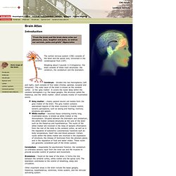
Weighing about 3 pounds (1.4 kilograms), the brain consists of three main structures: the cerebrum, the cerebellum and the brainstem. Cerebrum - divided into two hemispheres (left and right), each consists of four lobes (frontal, parietal, occipital and temporal). The outer layer of the brain is known as the cerebral cortex or the ‘grey matter’. It covers the nuclei deep within the cerebral hemisphere e.g. the basal ganglia; the structure called the thalamus, and the ‘white matter’, which consists mostly of myelinated axons. – closely packed neuron cell bodies form the grey matter of the brain. Cerebellum – responsible for psychomotor function, the cerebellum co-ordinates sensory input from the inner ear and the muscles to provide accurate control of position and movement. Basal Ganglia Thalamus and Hypothalamus Ventricles Limbic System Reticular Activating System Neurons Glia.
The Brain Is Ready for Its Close-Up. How to Build a Human Brain, in 7 Easy Steps. Functional magnetic resonance imaging. Researcher checking fMRI images Functional magnetic resonance imaging or functional MRI (fMRI) is a functional neuroimaging procedure using MRI technology that measures brain activity by detecting associated changes in blood flow.[1] This technique relies on the fact that cerebral blood flow and neuronal activation are coupled.
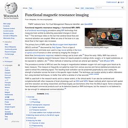
When an area of the brain is in use, blood flow to that region also increases. The primary form of fMRI uses the Blood-oxygen-level dependent (BOLD) contrast,[2] discovered by Seiji Ogawa. The procedure is similar to MRI but uses the change in magnetization between oxygen-rich and oxygen-poor blood as its basic measure. This measure is frequently corrupted by noise from various sources and hence statistical procedures are used to extract the underlying signal. FMRI is used both in the research world, and to a lesser extent, in the clinical world. Overview[edit] History[edit] Three studies in 1992 were the first to explore using the BOLD contrast in humans. Diffusion MRI.
Diffusion MRI (or dMRI) is a magnetic resonance imaging (MRI) method which came into existence in the mid-1980s.[1][2][3] It allows the mapping of the diffusion process of molecules, mainly water, in biological tissues, in vivo and non-invasively.
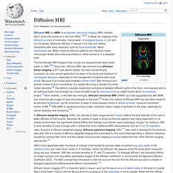
Molecular diffusion in tissues is not free, but reflects interactions with many obstacles, such as macromolecules, fibers, membranes, etc. Water molecule diffusion patterns can therefore reveal microscopic details about tissue architecture, either normal or in a diseased state. The first diffusion MRI images of the normal and diseased brain were made public in 1985.[4][5] Since then, diffusion MRI, also referred to as diffusion tensor imaging or DTI (see section below) has been extraordinarily successful. Its main clinical application has been in the study and treatment of neurological disorders, especially for the management of patients with acute stroke. Diffusion[edit] Given the concentration and flux where D is the diffusion coefficient. . Jonah Lehrer on the fMRI Scan: Little-Known Pitfalls.