
Brain connections last as long as the memories they store, Stanford neuroscientist finds. June 22, 2015 A team of Bio-X scientists applied microscopy know-how to a long-standing theory in neuroscience: if brain connections called synapses store memories, those synapses should last as long as the memories themselves.
It turns out they do, as Mark Schnitzer was able to show. By Amy Adams Stanford researchers have used microscopy tools that they developed in the lab to monitor the synapses between hippocampal neurons, opening a window onto the workings of short-term and long-term memory. (Creations / Shutterstock) Our memories are as fleeting as the brain structures that store them, or so the theory goes. The idea seemed good, but has been hard to test. Now Mark Schnitzer, an associate professor of biology and of applied physics, has leveraged microscopy tools developed in his lab and for the first time was able to monitor the connections, called synapses, between hippocampal neurons and confirm what neuroscientists thought might be happening. Mobile memories. Neuroimaging: Separating the Promise from the Pipe Dreams. Colorful brain images may tempt researchers to make claims that outpace solid scientific data—and may tempt the public to believe those claims.

In particular, although brain imaging has provided solid evidence of alterations in brain structures and functions associated with many psychiatric disorders, it can be used neither to diagnose such disorders nor to determine exactly how treatments work—at least not yet. Keeping some key ideas in mind can help us evaluate the next report of a brain-imaging “breakthrough.”
On any given day you are likely to see a news report mentioning brain imaging. As I write this, a quick search of recent news stories yields the following headlines: The hippocampus and the sense of smell. A review, by Alf Brodal. Brain 1947: 70; 179–222. Writing in 1587, Giulio Cesare Aranzi (1530–1589) describes a structure ‘continuous with the vaulted body or tortoise (fornix) which has an uneven or bent form that resembles the appearance of a hippocampus, that is a sea-horse’ (De humano foetu liber tertio editus, ac recognitus.
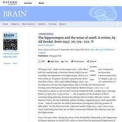
Euisdem anatomicarum observationum liber, Chapter 3, pp. 45). Paul Broca (1824–1880) and Ludwig Edinger (1855–1918) traced direct connections of the olfactory tract into the hippocampus. But in Trabajos del Laboratorio de Investigaciones Biologicas del la Universidad de Madrid (Tomo 1: 1901–02; 1–227. Many fibres from the olfactory epithelium terminate in the anterior olfactory nucleus, which lies posterior within the olfactory bulb, and pass from there (with others that do not relay in the anterior olfactory nucleus) to the piriform lobe cortex (Fig. 1).
Allegedly, connections exist between the two olfactory bulbs and both anterior olfactory nuclei through the anterior commissure. Figure 1 Figure 2 Figure 3. Olfaction and Memory. Olfaction and Memory Have you ever had the experience where you perceive a smell and suddenly remember an event that you'd forgotten for years?
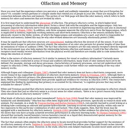
Or, consider certain perfume or cologne smells that remind you of people in your life. These are examples of the connection between olfaction and memory. This section of our Web page will describe odor memory, which refers to both memory for odors and memories that are evoked by odors. It is first important to understand the physiology of olfaction. It may be significant that olfactory neurons are unmyelinated, making olfaction the slowest of all the senses. What is the nature of olfactory memory?
Although Engen and colleagues (Engen, 1987) claimed that odor memory does not have a short-term component, more recent research has supported the existence of olfactory short-term memory (White & Treisman, 1997). White and Treisman posited that olfactory memory occurs because individuals assign verbal meanings to olfactory stimuli. Brain.swf (application/x-shockwave-flash Object) Brain Map. The brain has many parts including the cerebral cortex, brain stem and cerebellum.
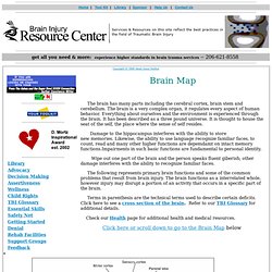
The brain is a very complex organ, it regulates every aspect of human behavior. Everything about ourselves and the environment is experienced through the brain. It has been described as a three pound universe. Brain Structures and Their Functions. The nervous system is your body's decision and communication center.
The central nervous system (CNS) is made of the brain and the spinal cord and the peripheral nervous system (PNS) is made of nerves. Together they control every part of your daily life, from breathing and blinking to helping you memorize facts for a test. Nerves reach from your brain to your face, ears, eyes, nose, and spinal cord... and from the spinal cord to the rest of your body. Sensory nerves gather information from the environment, send that info to the spinal cord, which then speed the message to the brain.
The brain then makes sense of that message and fires off a response. The brain is made of three main parts: the forebrain, midbrain, and hindbrain. C:\WRIT\PAPERS\EuroChap.wpd - basicneuro.pdf. Neuroanatomy Tutorial 9 (Brain) Interactive Neuroanatomy Atlas.