» Why Anatomy Is Important. I’ve had pain in to the left of my spine, a little inferior (below) the scapula and just lateral of the vertebrae, off and on over the last year.

It’s often not debilitating, but it will get worse when I sit for long periods of time. I have strained several times where I couldn’t train for a few sessions. I did it in January when I was playing Skyrim, and another time it happened when I was front squatting in Texas (2010, I think). The earliest I ever strained it was before I even did CrossFit, so that must have been in 2007 (I was doing a dumbbell row). I have a slight curve to my thoracic spine which would probably explain why the erector spinae muscles on that side have issues. I’ve tried a lot of different things from Mobility WOD, and they helped, but the pain still lingers. All of the above is to give you an idea of the data I’ve collected on this injury and how it could help me work on it to improve it.
I use this book every time someone asks me an injury question. Muscle. Muscle tissues are derived from the mesodermal layer of embryonic germ cells in a process known as myogenesis.
There are three types of muscle, skeletal or striated, cardiac, and smooth. Muscle action can be classified as being either voluntary or involuntary. Cardiac and smooth muscles contract without conscious thought and are termed involuntary, whereas the skeletal muscles contract upon command. [citation needed] Skeletal muscles in turn can be divided into fast and slow twitch fibers. Muscles are predominantly powered by the oxidation of fats and carbohydrates, but anaerobic chemical reactions are also used, particularly by fast twitch fibers.
Myofibril. A diagram of the structure of a myofibril Sliding filament model of muscle contraction A myofibril (also known as a muscle fibril) is a basic rod-like unit of a muscle.[1] Muscles are composed of tubular cells called myocytes, also known as muscle fibers, and these cells in turn contain many chains of myofibrils.

They are created during embryo development in a process known as myogenesis. Myofibrils are composed of long proteins such as actin, myosin, and titin, and other proteins that hold them together. These proteins are organized into thin filaments and thick filaments, which repeat along the length of the myofibril in sections called sarcomeres. Actomyosin motors are important in muscle contraction (relying in this case on "classical myosins") as well as other processes like retraction of membrane blebs, filiopod retraction, and uropodium advancement (relying in this case on "nonclassical myosins").
Structure[edit] Formation[edit] Appearance[edit] Action[edit] Myofibril.jpg (JPEG Image, 504 × 324 pixels) Actin. A large number of illnesses and diseases are caused by mutations in alleles of the genes that regulate the production of actin or of its associated proteins.
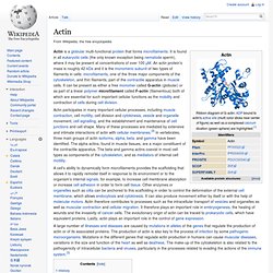
The production of actin is also key to the process of infection by some pathogenic microorganisms. Mutations in the different genes that regulate actin production in humans can cause muscular diseases, variations in the size and function of the heart as well as deafness. The make-up of the cytoskeleton is also related to the pathogenicity of intracellular bacteria and viruses, particularly in the processes related to evading the actions of the immune system.[3] History[edit] Actin was first observed experimentally in 1887 by W.D.
Myosin. Part of the myosin II structure.

Atoms in the heavy chain are colored red on the left-hand side, and atoms in the light chains are colored orange and yellow. Myosins comprise a family of ATP-dependent motor proteins and are best known for their role in muscle contraction and their involvement in a wide range of other eukaryotic motility processes. They are responsible for actin-based motility. The term was originally used to describe a group of similar ATPases found in striated and smooth muscle cells.[1] Following the discovery by Pollard and Korn of enzymes with myosin-like function in Acanthamoeba castellanii, a large number of divergent myosin genes have been discovered throughout eukaryotes.
Thus, although myosin was originally thought to be restricted to muscle cells (hence, "myo"), there is no single "myosin" but rather a huge superfamily of genes whose protein products share the basic properties of actin binding, ATP hydrolysis (ATPase enzyme activity), and force transduction. Titin. Endomysium. The endomysium, meaning within the muscle, is a wispy layer of areolar connective tissue that ensheaths each individual muscle fiber.

It also contains capillaries and nerves. It overlies the muscle fiber's cell membrane: the sarcolemma. The term cardiac skeleton is sometimes considered synonymous with endomysium[clarification needed], but sometimes cardiac skeleton refers to the combination of the endomysium and perimysium. Anti-endomysial antibodies (EMA) are present in celiac disease. Sarcolemma. The sarcolemma (Sarco (from Sarx) from Greek; Flesh, and Lemma from Greek; sheath.) also called the myolemma, is the cell membrane of a striated muscle fiber cell.[1] It consists of a plasma membrane which is a lipid bi-layer, and an outer coat consisting of a thin layer of polysaccharide material (glycocalyx) that contacts the Basement membrane containing numerous thin collagen fibrils and specialized proteins such as Laminin[2] to provide a scaffold for the muscle fiber to adhere to.

Muscular System Part 1 NSCA Certification. Muscular System Part 2 NSCA Certification.
Shoulder.