
CLINICAL TRIALS - VERY GOOD - Phase I Study of a Dendritic Cell Vaccine for Patients With Either Newly Diagnosed or Recurrent Glioblastoma. Understanding Dopaminergic Cell Death Pathways in Parkinson Disease. Review 1 Inserm, U 1127, F-75013 Paris, France2 CNRS, UMR 7225, F-75013 Paris, France3 Sorbonne Universités, UPMC Univ Paris 06, UMRS 1127, F-75013 Paris, France4 Institut du Cerveau et de la Moelle Epinière, ICM, F-75013 Paris, France Available online 18 May 2016 Choose an option to locate/access this article: Check if you have access through your login credentials or your institution Check access doi:10.1016/j.neuron.2016.03.038 Get rights and content Parkinson disease (PD) is a multifactorial neurodegenerative disorder, the etiology of which remains largely unknown.
. © 2016 Elsevier Inc. Clinical Gate. Simple, Scalable Proteomic Imaging for High-Dimensional Profiling of Intact Systems. Figure 3 SWITCH Enables Proteomic Imaging and High-Dimensional Quantitative Phenotyping of Human Clinical Samples (A) ROI from Figure 2 E showing semi-automatically detected locations and sizes of blood vessels (lectin) and diverse cell types (GFAP+, NeuN+, SMI-32+, CB+, CR+, PV+) in human visual cortex.
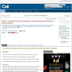
The identified objects are overlaid on maximum intensity-projections of raw images of the corresponding channels (dark gray). Dashed lines divide cortical layers I–VI. (B) 3D rendering of the boxed region in (A) (200 μm wide × 200 μm high × 104 μm deep) showing identified cells and blood vessels. (C) A heat map of the soma size distribution of NeuN+ cells, showing bimodal peaks at cortical layers III and V. (D) Density profiles of various cell types. (E) Comparison of cell sizes among different types of cells. (F) Distribution of neurons expressing various subsets of calcium-binding proteins in the human visual cortex. (G) A representative NeuN+/SMI-32+/CB+/PV+ cell.
A Tumor Suppressor Function for Notch Signaling in Forebrain Tumor Subtypes. Article 1 Department of Biomedicine, University of Basel, Mattenstrasse 28, 4058 Basel, Switzerland2 Department of Biomedicine, University Hospital Basel, Spitalstrasse 21, 4031 Basel, Switzerland3 Department of Cellular and Molecular Medicine, Lerner Research Institute, Cleveland Clinic, 9500 Euclid Avenue, NC 10, Cleveland, OH 44195, USA4 Department of Biomedicine, University of Basel, Kingelbergstrasse 50-70, 4056 Basel, Switzerland5 Division of Neuropathology, Institute of Pathology, University of Basel, Schoenbeinstrasse 40, 4031 Basel, Switzerland6 Division of Pediatric Neurooncology, German Cancer Research Center (DKFZ), 69121 Heidelberg, Germany Received 27 April 2015, Revised 6 August 2015, Accepted 16 October 2015, Available online 5 December 2015 Published: December 3, 2015 Choose an option to locate/access this article: Check if you have access through your login credentials or your institution Check access doi:10.1016/j.ccell.2015.10.008 Get rights and content Highlights Summary.
USP Standards. Sparrho. Sparrho. NMDA and AMPA Receptors. Map Viewer 1.0 (beta) Map Viewer 1.0 (beta) Map description This cortico-cortical map provides an overview of projection patterns for each cortical injection case available in iConnectome.
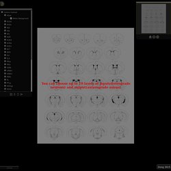
Data were manually reconstructed from raw images--axons appear as shaded regions and retrogradely labeled cells appear as colored dots. A large dot indicates injection site. Main view To navigate within image: drag image within main window or drag zone within the navigation window or click an area within navigation windowTo zoom in: double click with the mouse or use the mouse scroll wheel or simply press the key of the navigation windowTo zoom out: double click with the mouse or use the mouse wheel or press the key of the navigation window Tree view menu window Navigation window Miscellaneous To measure with scale: drag and move the scale on top of the target locationTo show help: click on the map viewer button placed on the upper leftTo show quick help: mouse over to the toolbar of the tree view menu or the navigator.
Understanding the Brain: The Neurobiology of Everyday Life - The University of Chicago. About the Course The Neurobiology of Everyday Life is a 10-week course intended for anyone interested in how the nervous system works.

The course starts by introducing basic neuroanatomy, neurodevelopment and mechanisms of neural communication. Students will gain an understanding of how injury and disease of different types and in different locations can alter a person’s life. Students will then use their understanding of fundamental neuroanatomy and physiology to examine how: 1) we perceive the outside world; 2) we act in the world either volitionally or emotionally; 3) our nervous system allows us to live; and 4) cognition operates to make us the human individuals that we are. We will look at the neurobiology of everyday situations such as multitasking (walking and chewing gum) and at the ways in which the nervous system commonly fails us (e.g. motion sickness).
If you'd like to get started thinking about the brain now, follow us on Twitter @neuromooc! Course Syllabus 1. 2. 3. 4. 5. 6. Home.