
Sm.stanford. Vidéos tuto - Samsung HS40 sur Vimeo. Build your clinical skills and become an expert. Medical Books Free Download Pdf [jen suis page 43) Radiology » Free PDF EPUB Medical Books. Pulmonary Embolism — Ultrasound Idiots. Carotid and Vertebral Arteries.
Guidelines. Trucs rigolos, goodies, images.. Articles, études sur écho et MG. Scrotal ultrasound - Startradiology. Indication/Technique Scrotal ultrasound is requested when pathology is suspected.
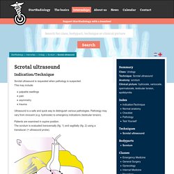
This may include: palpable swellings pain asymmetry trauma Ultrasound is a safe and quick way to distinguish various pathologies. Pathology may vary from innocent (e.g. hydrocele) to emergency indications (testicular torsion). Patients are examined in supine position. Figure 1. Figure 2. By moving and rotating the transducer, each part of the scrotum is assessed systematically. In general, scrotal ultrasound (as other ultrasound examinations) are performed in the transversal plane (fig. 3): the top of the ultrasound image is the anterior side; the bottom is the posterior side.
Figure 3. As a general rule, in ultrasound in the sagittal plane (fig. 4): the top of the ultrasound image is the anterior side and the bottom is the posterior side. right on the image is the foot side (= caudal) and left is the head side (= cranial).
Cours divers. Echo abdo. Musculo-tendineux. Gynéco. Echo pulm. Urgences. Vasculaire. Canadian Point of Care Ultrasound Society. EmDOCs.net – Emergency Medicine EducationUltrasound G.E.L. Archives - emDOCs.net - Emergency Medicine Education. Clinical Ultrasonography 101: Seeing Clearer with Ocular Ultrasound - CanadiEM.
Ever feel frustrated with your eye exam?
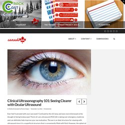
Confused by the slit lamp and even more distressed at the thought of doing funduscopy? Point-of-care ultrasound (POCUS) is taking over emergency medicine and can definitely help improve your eye evaluation. The eye is an ideal structure for viewing with ultrasound since it is a superficial structure that is conveniently filled with fluid. However, the spherical three-dimensional structure of the ocular globe can be confusing when trying to view it using a two-dimensional ultrasound image. This POCUS post offers an introduction to ocular ultrasound and some tips to make sure you capture every detail. Ocular Ultrasound Applications Ocular ultrasound has a range of clinical applications:
Smacc sonowars. Fantomes et simulateurs. Modeles anatomiques. Unlocking the Value of Point of Care Ultrasound in Internal Medicine. SonoSlam 2017. If you attended the AIUM convention the past 2 years you may have heard mention of SonoSlam in passing.
So what is it? SonoSlam is a medical student ultrasound competition and educational event. It was conceived as an idea to promote medical student ultrasound and was officially born in Orlando in 2015. A few members of the medical education committee were discussing how to get students more engaged in ultrasound at the national level. A national ultrasound student interest group had been formed and got behind the idea of nationalizing ultrasound activities for medical students. For more information about SonoSlam or if you are interested getting involved please email us: sonoslam@gmail.com. Written by Creagh Boulger, Rachel Liu, and Dave Bahner. Posakony notes on the Howry scanners. Historical Notes from Mr.
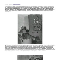
Gerald Posakony: I first started working with Dr. Douglass Howry in 1949 when he moved to Denver to work with Roderick Bliss in a company called Decimeter, Inc. Rod Bliss had an excellent background in electronics and microwaves and Howry relied on him to help him understand what conceptually could be done in the field of ultrasonics that might be analogous to the field of microwaves and useful to the medical diagnostic field. Howry was an avid reader and his mind was going around constantly. The picture shows the original reduction to practice of Howry's "Somascope". Échographie : WebSite Médecine tactique CITERA 69 - Baron Desgenettes. Press GM et Al.
J Emerg Med. 2014 Dec;47(6):638-45 L'apport de l'échographie est incontournable pour la prise en charge des traumatisés. Son emploi en prehospitalier est proposé. Pour autant la mise en oeuvre de ce moyen d'exploration n'est pas si simple et demande une grande expertise. Le travail présenté porte sur la mise en oeuvre de ce type d'exploration par technicinens paramédicaux expérimentés et ayant suivi une formation sur une période de deux mois. malgré cela leur performance reste modeste. 5 Min Sono Vids - 5 minute sono. Foie Archives - PinkyBone. INDICATION Bilan RÉSULTATS Foie de taille normale, d’échostructure homogène et contours réguliers.
Foie dysmorphique (hypertrophie du I, atrophie du IV), de contours irréguliers et d’échostructure grossière, nodulaire. Tronc porte, branches portes intra-hépatiques perméables, de sens physiologique. Veines hépatiques de calibre habituel, perméables, bien modulées en Doppler pulsé. BIENVENUE SUR LE SITE DE WINFOCUS-FRANCE. Journal of Ultrasound in Medicine. SOS Echographie. Thyroïde [échographie] - PinkyBone.