The Common Bile Duct. Renal ultrasound video 1, University of Florida Nephrology by Dr. Koratala (Twitter:@NephroP) FOAMed - Renal Fellow Network. NephJC is an online Twitter based journal club which regularly has nephrologists, residents, fellows, cardiologists, internists, urologists, radiologists, pharmacologists, and patients contribute to the discussion.

NephJC has a static webpage, a newsletter, a Twitter feed, and a Facebook page. Neph Sim is a mobile-friendly teaching tool that can be used to learn or teach how to establish a differential diagnosis, understand pathophysiology, and review urinalysis, ultrasound, kidney pathology, vascular access, kidney transplantation, acid-base physiology, electrolyte disturbances, and more. NephroPOCUS is a comprehensive resource for point of care ultrasound in nephrology (POCUN) GlomCon is a new project with the goal to create enabling platforms for clinicians and scientists to exchange ideas, participate in online conferences, and collaborate on basic science and clinical research projects WashU Nephrology Web Episodes are monthly videos produced by Tim Yau.
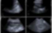
Hepatobiliary — TPA. Kyste Archives - PinkyBone. INDICATION Bilan de RESULTATSRein droit, Bonne différenciation parenchymato-sinusale.
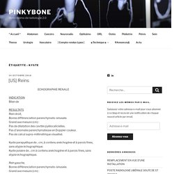
Grand axe mesure (cm) : Pas de dilatation des cavités pyélocalicielles. Pas d’anomalie parenchymateuse en Doppler couleur. Pas de calcul supra-millimétrique visualisé. Kyste parapyélique de .. cm, à contenu anéchogène et à parois fines, sans atypie échographique. Rein gauche, Bonne différenciation parenchymato-sinusale. Vessie en bonne réplétion, à parois fines et à contenu anéchogène. CONCLUSIONPas de dilatation des cavités pyélocalicielles.Absence de signe échographique d’infection. Kystes rénaux. Kyste Archives - PinkyBone. Pancreas carcinoma cancer ultrasonography gallbladder sludje main biliary duct dilatation wirsung choledoc cancer du pancréas dilation voie biliaire intra hépatique extra cholédoque boue biliaire vésicule échographie tumeur pancréas cancer doppler echogra. Introduction : Carcinoma of the pancreas is a highly malignant tumor with poor prognosis (The overall 5-year survival rate for this disease is less than 5%).
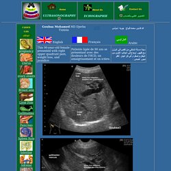
It represents 95% of all malignant pancreatic tumors. 60% are round in the head of the pancreas, 20 per cent in the body or rail of the pancreas and 20 pet cent involve the whole pancreas. About 80% of tumors in the pancreas are focal. It primarily occurs in the sixth to eighth decades of life with a male preponderance. The risk factors include alcohol abuse, diabetes, smoking , asbestos exposure, hereditary pancreatitis and chronic calcific pancreatitis. Histologically 95 pet cent of pancreatic carcinomes are adenocarcinoma, which usually originale from ductal elements. Pathophysiology: Pancreatic cancers can arise from both the exocrine and endocrine portions of the pancreas. Ultrasound : The detection rate by any imaging procedure depends on an adequately visualized gland. 04 Peritoneum. Hot Tips - Locating the Common Bile Duct with Ultrasound. Ultrasound Video showing an inflamed appendix. QA video of appendicitis. Ultrasound Teaching Manual Transducer Positioning and Resulting Ultrasound Images Liver and Gall.
Ultrasound Teaching Manual Transducer Positioning and Resulting Ultrasound Images Liver and Gall. Small bowel obstruction: Diagnosis by ultrasonography. A 64 year old man with an extensive history of abdominal surgeries presents to the emergency department with abdominal pain and vomiting.
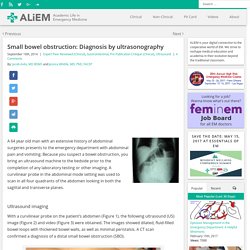
Because you suspect a bowel obstruction, you bring an ultrasound machine to the bedside prior to the completion of any laboratory testing or other imaging. A curvilinear probe in the abdominal mode setting was used to scan in all four quadrants of the abdomen looking in both the sagittal and transverse planes. Ultrasound imaging With a curvilinear probe on the patient’s abdomen (Figure 1), the following ultrasound (US) image (Figure 2) and video (Figure 3) were obtained. The images showed dilated, fluid-filled bowel loops with thickened bowel walls, as well as minimal peristalsis. Figure 1. Figure 2. Figure 3. SBO: Background Annually, in the United States, approximately 10% of all visits to the emergency department, or 13 million patients, present with the chief complaint of abdominal pain1. SBO: History and Physical SBO: Imaging. Ultrasound of the Pancreas. Acute Abdomen - Role of Ultrasound.
The diagnosis of sigmoid diverticulitis is often made on clinical grounds.
In the classical case the patient presents with localized pain and guarding in the left lower abdomen, fever, leukocytosis and, later on, elevation of the sedimentation rate. However, the diagnosis is not always clear. On one hand the clinical signs may be so atypical that initially another diagnosis is considered, as urinary tract infection, renal colic, perforated peptic ulcer, adnexitis or, -in case of diverticulitis in a rightsided loop of sigmoid- appendicitis.
On the other hand, the clinician may think of sigmoid diverticulitis while in fact another condition is present, as sigmoid carcinoma, epiploic appendagitis, a gynecological or urological condition or even a ruptured aortic aneurysm. In all of these cases, US may play a role by making the correct diagnosis at an early point in time. Pleased to meet you... Check out these Rolling Stones! @ACEP_EUS @UltrasoundJelly @ultrasoundpod @DavidPigottMD #FOAMus #UABWOTW.