
How the Brain Stops Time. One of the strangest side-effects of intense fear is time dilation, the apparent slowing-down of time. It's a common trope in movies and TV shows, like the memorable scene from The Matrix in which time slows down so dramatically that bullets fired at the hero seem to move at a walking pace. In real life, our perceptions aren't keyed up quite that dramatically, but survivors of life-and-death situations often report that things seem to take longer to happen, objects fall more slowly, and they're capable of complex thoughts in what would normally be the blink of an eye. Now a research team from Israel reports that not only does time slow down, but that it slows down more for some than for others.
Anxious people, they found, experience greater time dilation in response to the same threat stimuli. Somatic markers hypothesis. The somatic marker hypothesis (SMH) proposes a mechanism by which emotional processes can guide (or bias) behavior, particularly decision-making.[1][2] This hypothesis has been formulated by Antonio Damasio. Hypothesis[edit] When individuals make decisions, they must assess the incentive value of the choices available to them, using cognitive and emotional processes. When the individuals face complex and conflicting choices, they may be unable to decide using only cognitive processes, which may become overloaded. The amygdala and VMPFC are essential components of this hypothesized mechanism and therefore damage to either structure will disrupt their proposed action in mediating the development and action of somatic markers.
A major source of supporting evidence for this theory is provided by experiments using the Iowa gambling task.[5] Research background[edit] VMPFC patients also have difficulty expressing and experiencing appropriate emotions. Working mechanism[edit] Experiments[edit] Feelings of Knowing : The Frontal Cortex. Clive Thompson has a wonderful article in the NY Times Magazine on Watson, the supercomputer programmed to excel at Jeopardy.
Thompson delves into the clever heuristics used to generate singular answers to ambiguous questions. (Watson relies on massive amounts of parallel processing, so that “he” is running thousands of Google searches simultaneously.) While Watson’s performance is certainly impressive, I thought the most interesting part of the story involved the failings of the machine. It’s easy to rhapsodize about the ever escalating speed of microchips, but it turns out that Watson is often too slow at ringing the buzzer: In more than 20 games I witnessed between Watson and former “Jeopardy!”
This anecdote highlights one of the most impressive talents of the human mind. What’s interesting about this mental hiccup is that, even though the mind can’t remember the information, it’s convinced that it knows it. This is where feelings of knowing prove essential. Brain. Cortex Command. Visuospatial ability: Studies.
A Neuroscientist Uncovers A Dark Secret. THE AMYGDALA AND THE EMOTIONS. Chapter 9 — The Amygdala and the Emotions by Ben Best This installment is something of a digression in my "systematic" attempt to investigate the anatomical basis of mind.
Here I investigate in detail a single component of the "Limbic System": the amygdala. Your Brain in Love. Men and women can now thank a dozen brain regions for their romantic fervor.
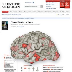
Researchers have revealed the fonts of desire by comparing functional MRI studies of people who indicated they were experiencing passionate love, maternal love or unconditional love. Together, the regions release neurotransmitters and other chemicals in the brain and blood that prompt greater euphoric sensations such as attraction and pleasure. Conversely, psychiatrists might someday help individuals who become dangerously depressed after a heartbreak by adjusting those chemicals.
Passion also heightens several cognitive functions, as the brain regions and chemicals surge. “It’s all about how that network interacts,” says Stephanie Ortigue, an assistant professor of psychology at Syracuse University, who led the study. Graphics by James W. Etymology of Neuroscience Terms. Technology Review: Brain Coprocessors. Ed Boyden, an Assistant Professor, Biological Engineering, and Brain and Cognitive Sciences at the MIT Media Lab, will give a presentation on using light to study and treat brain disorders at 3.30pm on Wednesday at EmTech 2010.
Watch a live feed of the session here. The last few decades have seen a surge of invention of technologies that enable the observation or perturbation of information in the brain. Functional MRI, which measures blood flow changes associated with brain activity, is being explored for purposes as diverse as lie detection, prediction of human decision making, and assessment of language recovery after stroke. Implanted electrical stimulators, which enable control of neural circuit activity, are borne by hundreds of thousands of people to treat conditions such as deafness, Parkinson’s disease, and obsessive-compulsive disorder. Neuro-links - Brain, Self and Society - LSE. BIOS now closed The BIOS Centre closed as a research centre of the School on 31 December 2011.

The BIOS research group is moving to King's College London as of January 2012 to become the research arm of a new Department of Social Science, Health and Medicine. Nikolas Rose will also be leaving the LSE at that time to become Head of this new Department. Further information and resources Research programmes and other activities previously undertaken in the BIOS Centre: Neuropsychological Assessment, Diagnostics and Cognitive Rehabilitation Therapy. Functional Neuroanatomy- Free Online Course. Wilderness Survival, Tracking, Nature, Wilderness Mind. Forge Survival Supply, The Gear Store For Survivalists.