
Soleus muscle. Structure[edit] The soleus is located in the superficial posterior compartment of the leg.
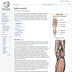
Not all mammals have a soleus muscle; one familiar species that lacks the soleus is the dog. Soleus is vestigial in the horse.[1] The soleus exhibits significant morphological differences across species. It is unipennate in many species. Gastrocnemius muscle. In humans, the gastrocnemius muscle (/ˌɡæstrɒkˈniːmiəs/ or /ˌɡæstrəkˈniːmiəs/; Latin, from Greek γαστήρ "stomach" and κνήμη (knēmē) "leg"; meaning "stomach of leg", referring to the bulging shape of the calf) is a very powerful superficial bipennate muscle that is in the back part of the lower leg.
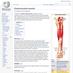
It runs from its two heads just above the knee to the heel. Structure[edit] The gastrocnemius is located with the soleus in the posterior (back) compartment of the leg. The lateral head originates from the lateral condyle of the femur, while the medial head originates from the medial condyle of the femur. Its other end forms a common tendon with the soleus muscle; this tendon is known as the calcaneal tendon or Achilles Tendon and inserts onto the posterior surface of the calcaneus, or heel bone. Flexor digitorum longus muscle. The flexor digitorum longus is situated on the tibial side of the leg.
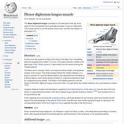
At its origin it is thin and pointed, but it gradually increases in size as it descends. This muscle serves to curl the second, third, fourth, and fifth toes (flexion of phalanges II-V). Vastus intermedius muscle. The vastus intermedius (/ˈvæstəs ˌɪntərˈmiːdi.əs/) (Cruraeus) arises from the front and lateral surfaces of the body of the femur in its upper two-thirds, sitting under the rectus femoris muscle and from the lower part of the lateral intermuscular septum.
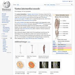
Its fibers end in a superficial aponeurosis, which forms the deep part of the quadriceps femoris tendon. The vastus medialis and vastus intermedius appear to be inseparably united, but when the rectus femoris has been reflected[when defined as?] Rectus femoris muscle. The rectus femoris muscle (/ˈrɛktəs ˈfɛmərɨs/) is one of the four quadriceps muscles of the human body.
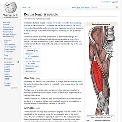
The others are the vastus medialis, the vastus intermedius (deep to the rectus femoris), and the vastus lateralis. All four parts of the quadriceps muscle attach to the patella (knee cap) via the quadriceps tendon. The rectus femoris is situated in the middle of the front of the thigh; it is fusiform in shape, and its superficial fibers are arranged in a bipenniform manner, the deep fibers running straight down to the deep aponeurosis. Its functions are to flex the thigh at the hip joint and to extend the leg at the knee joint.[1] Biceps brachii muscle. In human anatomy, the biceps brachii (/ˈbaɪsɛps ˈbræki.aɪ/), commonly known as the biceps, is a two-headed muscle that lies on the upper arm between the shoulder and the elbow.
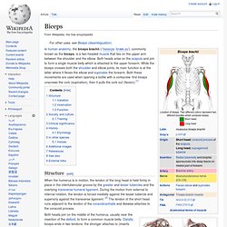
Both heads arise on the scapula and join to form a single muscle belly which is attached to the upper forearm. While the biceps crosses both the shoulder and elbow joints, its main function is at the latter where it flexes the elbow and supinates the forearm. Both these movements are used when opening a bottle with a corkscrew: first biceps unscrews the cork (supination), then it pulls the cork out (flexion).[1] Tensor fasciae latae muscle. The tensor fasciae latae (or tensor fasciæ latæ) (/ˈtɛnsər ˈfæʃi.iː ˈleɪtiː/) is a muscle of the thigh.
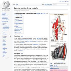
Structure[edit] Gluteus maximus muscle. The gluteus maximus (also known as glutæus maximus or, collectively with the gluteus medius and minimus, the glutes) is the largest and most superficial of the three gluteal muscles.
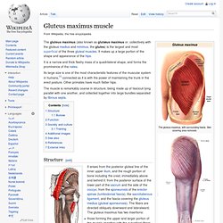
It makes up a large portion of the shape and appearance of the hips. It is a narrow and thick fleshy mass of a quadrilateral shape, and forms the prominence of the nates. Its large size is one of the most characteristic features of the muscular system in humans,[1] connected as it is with the power of maintaining the trunk in the erect posture. Other primates have much flatter hips. The muscle is remarkably coarse in structure, being made up of fasciculi lying parallel with one another, and collected together into large bundles separated by fibrous septa. Structure[edit] Muscles of the gluteal and posterior femoral regions, showing origin and insertion of gluteus maximus muscle. Bursae[edit] Palmaris longus muscle. The palmaris longus is seen as a small tendon between the flexor carpi radialis and the flexor carpi ulnaris, although it is not always present.
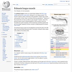
The muscle is absent in about 14 percent of the population, however this number varies greatly in African, Asian, and Native American populations.[1] Absence of this muscle does not have an effect on grip strength;[2] however, lack of the palmaris longus muscle is disadvantageous in that its tendon cannot be harvested as a graft if necessary. Some evolutionary scientists maintain that as it is no longer needed by humans, evolution is dispensing with the tendon. [citation needed] Brachioradialis. Structure[edit] Distal end of Humerus/styloid process of radius Innervation[edit]
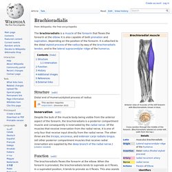
Triceps brachii muscle. Structure[edit] Biceps femoris muscle. The biceps femoris (/ˈbaɪsɛps ˈfɛmərɨs/) is a muscle of the posterior (the back) thigh. As its name implies, it has two parts, one of which (the long head) forms part of the hamstrings muscle group. Structure[edit] It has two heads of origin; Infraspinatus muscle. In human anatomy, the infraspinatus muscle is a thick triangular muscle, which occupies the chief part of the infraspinatous fossa.[1] As one of the four muscles of the rotator cuff, the main function of the infraspinatus is to externally rotate the humerus and stabilize the shoulder joint. Structure[edit] It attaches medially to the infraspinous fossa of the scapula and laterally to the middle facet of the greater tubercle of the humerus.
Supraspinatus muscle. Structure[edit] The supraspinatus muscle arises from the supraspinous fossa, a shallow depression in the body of the scapular above its spine. The supraspinatus muscle tendon passes laterally beneath the cover of the acromion. Research in 1996 showed that the postero-lateral origin was more lateral than classically described.[1][2] Teres major muscle. The teres major muscle (Latin teres meaning 'rounded') is a muscle of the upper limb and one of seven scapulohumeral muscles.
It is a thick but somewhat flattened muscle, innervated by the lower subscapular nerve (C5 and C6). Structure[edit] Deltoid muscle. In human anatomy, the deltoid muscle is the muscle forming the rounded contour of the shoulder. Anatomically, it appears to be made up of three distinct sets of fibers though electromyography suggests that it consists of at least seven groups that can be independently coordinated by the central nervous system.[1] A study of 30 shoulders revealed an average mass of 191.9 grams (6.77 oz) (range 84 grams (3.0 oz)–366 grams (12.9 oz)) in humans.[2] Structure[edit] The deltoid originates in three distinct sets of fibers, often referred to as "heads":[3] Fick[14] divided these three groups of fibers, often referred to as parts (Latin: pars) or bands, into seven functional components:[15] the anterior part has two components (I and II); the lateral one (III); and the posterior four (IV, V, VI, and VII) components.
Side. Front. Back. Rectus abdominis muscle. Abdominal external oblique muscle. Thoracic diaphragm. The term "diaphragm" in anatomy can refer to other flat structures such as the urogenital diaphragm or pelvic diaphragm, but "the diaphragm" generally refers to the thoracic diaphragm. Trapezius muscle. Latissimus dorsi muscle. Pectoralis major muscle. The pectoralis major (/ˌpɛktəˈreɪlɨs ˈmeɪdʒər/) (from Latin: pectus, breast) is a thick, fan-shaped muscle, situated at the chest (anterior) of the human body.
Internal intercostal muscles. External intercostal muscles. The Intercostales externi (External intercostals) are eleven in number on either side. Zygomaticus major muscle. Sternocleidomastoid muscle. Orbicularis oculi muscle. Orbicularis oris muscle. Masseter muscle. Buccinator muscle.