
Clues to Autism Emerge from Brain Imaging. Action Points Two studies used imaging techniques to evaluate patients with autism spectrum disorder.One study using quantitative magnetic resonance imaging found that individuals with autism spectrum disorder have significant differences in cortical volume associated with separable variations in cortical thickness and surface area.Another study using positron emission tomography (PET) indicates excessive microglial activation in multiple brain regions in young adult subjects with autism spectrum disorder.
Young adults with autism spectrum disorder (ASD) had significant variation in cortical volume, thickness, and surface area compared with an age-matched control group, MRI brain scans showed. Differences in surface area accounted for a majority of the variation in cortical volume, but in almost a third of cases, neither cortical thickness nor surface area could account for the variation in volume. Most prior studies employed volumetric analyses. The study had some limitations. Autism brains are overly connected, studies find — Overly connected: Many pairs of brain regions — including those involved in sensory processing, emotion and motivation — are more tightly synchronized in children with autism (right) than in controls (left).
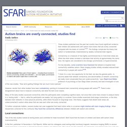
Three studies published over the past two months have found significant evidence that children and adolescents with autism have brains that are overly connected compared with the brains of controls1,2,3. The findings complicate the theory that autism is fundamentally characterized by weakly connected brain regions. Connectivity is a measure of how tightly synchronized two or more brain areas are. When two brain areas increase or decrease their activity at approximately the same time, the regions are considered to be strongly synchronized, or hyperconnected. For two decades, some scientists have theorized that altered or impaired brain connectivity underlies autism. Local connections: Two of the new studies looked at resting brains and controlled for head movement.
Electrical evidence: 1999_BCetal_FMRI.pdf. Functional MRI in Asperger Syndrome - Autism Research Centre. Mike Lombardo, Bhismadev Chakrabarti, Simon Baron-Cohen, Rosie Holt, Ed Bullmore, John Suckling, Meng-Chuan Lai, Amber Ruigrok, Richard Bethlehem, Dorothea Floris, Owen Parsons, EU-AIMS The ARC was the first group to show orbito-frontal and amygdala underactivity in the autistic brain using SPECT (single photon emission tomography) and later fMRI (functional magnetic resonance imaging) during empathy or 'mentalising' tasks.
These regions have been recognized to be part of the 'social brain', a network of regions that includes medial prefrontal cortex and the anterior cingulate, which do not function in the normal way in autism. We have also used fMRI during attentional tasks (such as the Embedded Figures Task in the visual modality or acoustic change-detection tasks in the auditory modality) to reveal the brain basis of the superior attention to detail in autism. The ARC was a collaborator on the MRC AIMS (Autism Imaging Multicentre Study) and is a collaborator on the EU-AIMS study. Center for Cognitive Brain Imaging - Welcome. How Brain Scans Can Diagnose Autism With 97% Accuracy.
Thinking about social interactions reveals brain patterns indicative of autism Right now, diagnosing disorders like autism relies heavily on interviews and behavioral observations.
But new research published in PLoS One shows that a much more objective measure—reading a person’s thoughts through an fMRI brain scan—might be able to diagnose autism with close to perfect accuracy. Lead study author Marcel Just, PhD, professor of psychology and director of the Center for Cognitive Brain Imaging at Carnegie Mellon University, and his team performed fMRI scans on 17 young adults with high-functioning autism and 17 people without autism while they thought about a range of different social interactions, like “hug,” “humiliate,” “kick” and “adore.” The researchers used machine-learning techniques to measure the activation in 135 tiny pieces of the brain, each the size of a peppercorn, and analyzed how the activation levels formed a pattern. Fmri+autism. Functional magnetic resonance imaging of autism spectrum disorders.
Functional MRI and autism, Functional Magnetic Resonance Imaging, fMRI and autism information from the Autism Center of Excellence, UC San Diego. We specialize in using functional magnetic resonance imaging (fMRI) to reveal the neural underpinnings of autism.
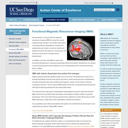
Our studies have been conducted with the participation of adults and adolescents with autism, as well as infants and toddlers at risk for the disorder. Structural MRI at the Autism Center of Excellence, UC San Diego. The Autism Center of Excellence has a long history of conducting studies aimed at better understanding brain structure and development in individuals with autism.
When autism was finally recognized as a biological disorder in the 1980s, ACE Director Eric Courchesne and his colleagues, were the first to show that the cerebellum is commonly observed to be abnormal in individuals with autism (see paper by Courchesne and colleagues, 1988). Courchesne and colleagues were also the first to identify brain growth abnormality in the first year of life in infants with autism. Currently, researchers at the Autism Center of Excellence are conducting MRI studies with children between 12 and 42 months using the sleep fMRI method. 'Autism discovery' - brain imaging reveals language development differences. Why do some children in the spectrum of autism disorder develop greater difficulties with language than others?
Discovering the neural basis of why some children with autism spectrum disorder (ASD) get better and develop good language and conversation abilities, and others do not, has been a research mystery of "utmost importance. " And the new findings, achieved through functional MRI (fMRI) brain imaging, open up new avenues for the causes and treatment of autism, say the researchers publishing in the journal Neuron.
The functional neuroimaging findings amount to a "discovery" on early language development in autism, say the authors in the research paper. Brain scans forecast language skills in autism — Center for Cognitive Brain Imaging - Welcome. How Brain Scans Can Diagnose Autism With 97% Accuracy. “The Nature of Brain Dysfunction in Autism: Functional Brain Imaging Studies” New brain scan to diagnose autism. Friday December 3 2010 The scans need testing in a wider range of autistic people “A foolproof test for autism in adults and children is “a major step” closer,” The Daily Telegraph has reported.
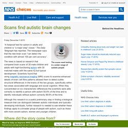
The newspaper says that the new brain scan “can detect the condition with almost 100 per cent accuracy”. Brain Scans May Predict Language Skills in Kids With Autism. But research is still preliminary, expert says WebMD News from HealthDay By Tara Haelle HealthDay Reporter THURSDAY, April 9, 2015 (HealthDay News) -- Sophisticated imaging tests could provide clues to how well a child with an autism spectrum disorder may develop language skills, possibly as early as when the child is just a year old, a new study suggests.
"We discovered that at the very first signs of autism in infants and toddlers, language-important brain regions already displayed striking differences between those who later had good versus poor language outcomes," said study co-author Eric Courchesne, a professor of neurosciences and co-director of the Autism Center of Excellence at the University of California, San Diego. UAB News - Research finds brain scans may aid in diagnosis of autism. AANE - Brain Imaging Studies in Autism Spectrum Disorders. Brain Imaging Studies in Autism Spectrum Disorders by Helen Tager-Flusberg, Ph.D., Boston University School of Medicine For the past ten years our research laboratory, now located at the Boston University School of Medicine, has been conducting brain imaging studies on children and adults with autism or Asperger Syndrome (referred to here as ASD) in collaboration with colleagues at Massachusetts General Hospital using magnetic resonance imaging, or MRI.
When we began there were very few published studies that had used this technology to investigate the brain structure and function in people with ASD; however in recent years there has been an huge increase in the literature in this area as more research groups take advantage of these exciting methods that allow us to investigate the brain in exquisite detail, in a painless non-invasive way.
We have been collecting structural MRI scans from children and adults with ASD ranging in age from 4 to 24 for quite a number of years now. What Is FMRI? - Center for Functional MRI - UC San Diego. Imaging Brain Activity Courtesy of Dr. David Shin, UC San Diego In your brain the activity of the neurons constantly fluctuates as you engage in different activities, from simple tasks like controlling your hand to reach out and pick up a cup of coffee to complex cognitive activities like understanding language in a conversation.
The brain also has many specialized parts, so that activities involving vision, hearing, touch, language, memory, etc. have different patterns of activity. Even when you rest quietly with your eyes closed the brain is still highly active, and the patterns of activity in this resting state are thought to reveal particular networks of areas that often act together.