
Tema 8: Intercambio pulmonar de gases. IMÁGENES Y ESQUEMAS DE LAS ARTIC. Articulación radiocarpiana.
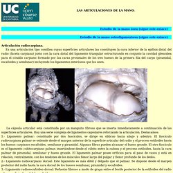
Es una articulación tipo condílea cuyas superficies articulares las constituyen la cara inferior de la epífisis distal del radio (faceta carpiana) junto con la cara distal del ligamento triangular estructurando en conjunto la cavidad glenoidea para el cóndilo carpiano formado por las caras proximales de los tres huesos de la primera fila del carpo (piramidal, escafoides y semilunar) incluyendo los ligamentos interóseos que los unen. La cápsula articular está constituida por un manguito fibroso que se inserta inmediatamente a continuación de las superficies articulares.
Hay una serie compleja de ligamentos capsulares reforzando la articulación. Destacamos: 1.- Ligamento palmar: constituido por dos fascículos, se dirige en oblicuo hacia abajo y adentro. 2.- Ligamento radiocarpiano dorsal: Este ligamento es más débil y delgado que el palmar. 4.- Ligamento colateral cubital del carpo: elástico y potente, en forma de abanico. ESQUEMA DE LA CIRCULACION CEREBRAL.
Making New Connections: How the Brain Can Develop Into Adulthood - Alice G. Walton - Life. A multi-year study has found that the brain's white matter -- fibers connecting cells -- can shift with employment, education, and relationships For many years, the convention was that after adolescence, the brain had done about as much developing as it was going to do.
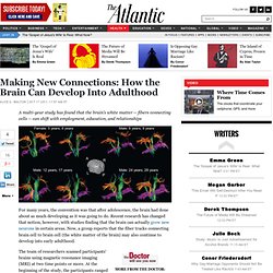
Recent research has changed that notion, however, with studies finding that the brain can actually grow new neurons in certain areas. Now, a group reports that the fiber tracks connecting brain cell to brain cell (the white matter of the brain) may also continue to develop into early adulthood. The team of researchers scanned participants' brains using magnetic resonance imaging (MRI) at two time points or more. At the beginning of the study, the participants ranged in age from 5 to 29, and the average gap between the first and second scanning was about four years. The frontal lobe is responsible for high-level executive function and attention. This article originally appeared on TheDoctorWillSeeYouNow.com. Brain Basics - 3D Model of Brain Injury. Animated Deceleration Injury from a Traumatic Brain Injury TBI Inform: Introduction to Brain Injury What Happens When a Brain Bleeds?

Areas of the Brain Affected by Concussion What is Chronic Traumatic Encephalopathy? Concussion Recovery The brain is incredibly complex. Located behind the forehead, the frontal lobes are the largest lobes of the brain. Planning organizing problem solving memory impulse control decision making selective attention controlling our behavior and emotions The left frontal lobe plays a large role in speech and language.
Problems After Injury Injury to the frontal lobes may affect: Cadaver Dissection Videos. Fundamentals of Neuroanatomy Video Lecture Course. THE AMYGDALA AND THE EMOTIONS. Chapter 9 — The Amygdala and the Emotions by Ben Best This installment is something of a digression in my "systematic" attempt to investigate the anatomical basis of mind.
Here I investigate in detail a single component of the "Limbic System": the amygdala. The basis of this investigation is a book of scientific papers entitled THE AMYGDALA, Edited by John P. Aggleton (Wiley-Liss, 1992). My motivation for this detailed study is the claim by Robert Ettinger that there exists in the brain a "self-circuit" that is the seat of feeling, and that this brain center is probably below the cerebrum because self&feeling are attributes of all animals.
The problem with the "Limbic System" is that is an abstraction, not an anatomical referent. (return to contents) The amygdala in a human is not much bigger than an almond (the Greek root word). (return to contents) Two major bundles of fibers connect the amygdala with other areas of the brain: the stria terminalis and the ventral amygdalofugal pathway. Science Visualized. Neuroscience. Med Resources. Main Page - Embryology. The Whole Brain Atlas.
Human anatomy.