GMO Human Embryos Have Already Been Created. A meeting at the FDA on experiments to create GMO humans has brought disturbing information to light.
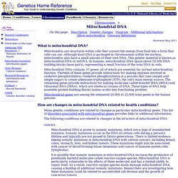
Action Alert! Today, the US Food and Drug Administration held day one of a public meeting outlining the creation of genetically modified humans. These experiments won’t take place in the distant future. In fact, GMO embryos have already been created via in vitro experiments. Specifically, the FDA is discussing the genetic manipulation of human eggs and embryos in order to prevent inherited mitochondrial disease and treat infertility. While the FDA has stated that the agency “recognizes” that there are “ethical and social policy issues” to be considered—and despite the fact that forty-four countries have already banned this kind of genetic manipulation—the FDA won’t bother to discuss if human clinical trials should take place (that’s considered to be “outside the scope” of the meeting). An FDA document outlines disturbing potential pitfalls of clinical trials to create GMO humans: Mitochondrial DNA. Electron microscopy reveals mitochondrial DNA in discrete foci.
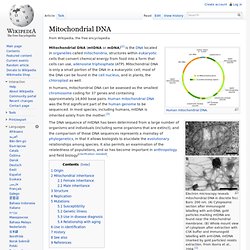
Bars: 200 nm. (A) Cytoplasmic section after immunogold labelling with anti-DNA; gold particles marking mtDNA are found near the mitochondrial membrane. What is mitochondrial DNA. Although most DNA is packaged in chromosomes within the nucleus, mitochondria also have a small amount of their own DNA.
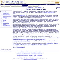
This genetic material is known as mitochondrial DNA or mtDNA. Mitochondria (illustration) are structures within cells that convert the energy from food into a form that cells can use. Each cell contains hundreds to thousands of mitochondria, which are located in the fluid that surrounds the nucleus (the cytoplasm). Mitochondria produce energy through a process called oxidative phosphorylation.
This process uses oxygen and simple sugars to create adenosine triphosphate (ATP), the cell’s main energy source. In addition to energy production, mitochondria play a role in several other cellular activities. Mitochondrial DNA contains 37 genes, all of which are essential for normal mitochondrial function. Molecular Expressions, a web site from the Florida State University Research Foundation, offers an illustrated introduction to mitochondria and mitochondrial .
Mitochondrial DNA. Many genetic conditions are related to changes in particular mitochondrial genes.
This list of disorders associated with mitochondrial genes provides links to additional information. The following conditions are related to changes in the structure of mitochondrial DNA. cancers Mitochondrial DNA is prone to somatic mutations, which are a type of noninherited mutation. Somatic mutations occur in the DNA of certain cells during a person's lifetime and typically are not passed to future generations.
Paternal mtDNA transmission. In genetics, paternal mtDNA transmission and paternal mtDNA inheritance refer to the incidence of mitochondrial DNA (mtDNA) being passed from a father to his offspring.
Eumetazoa. Eumetazoa (Greek: εὖ [eu], well + μετά [metá], after + ζῷον [zóon], animal) is a clade comprising all major animal groups except sponges, placozoa, and several other obscure or extinct life forms, such as Dickinsonia.
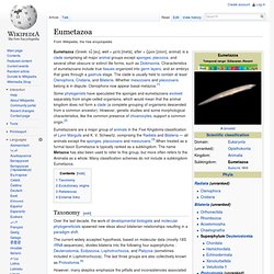
Characteristics of eumetazoans include true tissues organized into germ layers, and an embryo that goes through a gastrula stage. The clade is usually held to contain at least Ctenophora, Cnidaria, and Bilateria. Whether mesozoans and placozoans belong is in dispute. Ctenophora now appear basal metazoa.[1] Some phylogenists have speculated the sponges and eumetazoans evolved separately from single-celled organisms, which would mean that the animal kingdom does not form a clade (a complete grouping of organisms descended from a common ancestor). Eumetazoans are a major group of animals in the Five Kingdoms classification of Lynn Margulis and K. Taxonomy[edit] However, many skeptics emphasize the pitfalls and inconsistencies associated with the new data. Mitochondrion. Two mitochondria from mammalian lung tissue displaying their matrix and membranes as shown by electron microscopy History[edit] The first observations of intracellular structures that probably represent mitochondria were published in the 1840s.[13] Richard Altmann, in 1894, established them as cell organelles and called them "bioblasts".[13] The term "mitochondria" itself was coined by Carl Benda in 1898.[13] Leonor Michaelis discovered that Janus green can be used as a supravital stain for mitochondria in 1900.
Friedrich Meves, in 1904, made the first recorded observation of mitochondria in plants (Nymphaea alba)[13][14] and in 1908, along with Claudius Regaud, suggested that they contain proteins and lipids. Benjamin F. Kingsbury, in 1912, first related them with cell respiration, but almost exclusively based on morphological observations.[13] In 1913 particles from extracts of guinea-pig liver were linked to respiration by Otto Heinrich Warburg, which he called "grana".
Cristae[edit] Eukaryote. Eukaryotes can reproduce both asexually through mitosis and sexually through meiosis and gamete fusion.
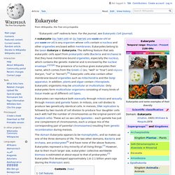
In mitosis, one cell divides to produce two genetically identical cells. In meiosis, DNA replication is followed by two rounds of cell division to produce four daughter cells each with half the number of chromosomes as the original parent cell (haploid cells). These act as sex cells (gametes – each gamete has just one complement of chromosomes, each a unique mix of the corresponding pair of parental chromosomes) resulting from genetic recombination during meiosis. Organelle.