
A framework for rapid prototyping of MR pulse sequences. The Medipix4 Collaboration. Kompetenz in medizinischer Bildgebung. Project WAKE-UP - Medical Imaging Research Institute (mediri) The mediri (medical imaging research institute) GmbH, founded in 2004, provides scientific research services in the field of medical imaging to customers from the academic community and pharmaceutical industry.
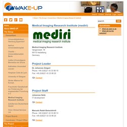
Main business areas of the mediri GmbH include the development of MR imaging methods, research in ultrasound imaging/therapy and medical image processing for clinical trial applications. Especially mediri’s services and offers of IT infrastructure developments for clinical trials are requested by global players in the pharmaceutical industry and major clinical trial research organisations. From these activities extensive knowledge has been built up in the development of infrastructure for image based clinical trials. This platform will provide the basis for the research conducted in this project.
Key Staff: Founding partner and current CEO of mediri is Prof. Dr. How to use DicomCleaner™ DicomCleaner™ is a free open source tool with a user interface for importing, "cleaning" and saving sets of DICOM instances (files).
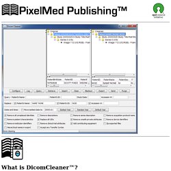
It can: Import files from a CD or the local hard drive or a shared network drive or any other media, with or without a DICOMDIRQuery and retrieve from remote DICOM devices"Clean" the DICOM "header" of a selected set of instancesBlackout burned in annotations in the pixel data of the cleaned filesExport the cleaned instances to a folder with a DICOMDIR, which can be burned to a CD or DVDSend the cleaned instances over the network to a remote DICOM device.
Nottingham Digestive Diseases Biomedical Research Centre. News - MR:comp GmbH. MRXCAT – Institute for Biomedical Engineering. Publications - k-Space Astronauts. Clinical PD & MR. Strategically Acquired Gradient Echo (STAGE) can image the whole brain and its vasculature in 9 minutes.
Features: * Co-registered T1W, PDW, T1 MAP, PD MAP, R2* MAP, SWI, tSWI, MRA and SWIM with a resolution of0.67mmx1.33mmx2.0mm and with 64 partitions. B1: TE=7.5ms/17.5ms, FA=6 degrees, 2.5 minutesB2: TE=8.75ms/18.75ms, FA=24 degrees, 2.5 minutesB3: TE=2.5ms/12.5ms (RP)/12.5MS (DP), FA=12 degrees, 4 minutes * Complemented by a T2 FLAIR image and by a DWI scan, a total of 12 minutes for the brain protocol.* Allows for separation of arteries and veins. * Allows for brain structure segmentation with homogeneous STAGE-enhanced T1W and QSM.* Data Processing done by SPIN software from MR Innovations. Yefeng Zheng. Yefeng Zheng Marginal Space Learning for Efficient Detection of 2D/3D Anatomical Structures in Medical Images Introduction Recently, machine learning based approaches have been successfully demonstrated on many 2D object detection problems (e.g., face detection, pedestrian detection, and vehicle detection in 2D images/video sequences).
In these methods, object detection or localization was formulated as a classification problem: whether an image block contains the target object or not. The robustness of the methods comes from the exhaustive search with the trained classifier during object detection on an input image. SimVascular. Downloads - CRIMSON. FAQ - Arterys. Do I need to download software to view, process and create a clinical report?

No. The Arterys™ interface is accessible via the Google Chrome web browser (we support the latest versions). No software plugin is needed. What kind of training do I need? The interface is intuitive and within 10 minutes you can feel comfortable with simple quantification. Purview ViVA - Cloud Medical Imaging Access and Storage. Questions and Answersin MRI - MRI Questions & Answers; MR imaging physics & technology. Associazione Italiana di Fisica Medica. OptiBET: optimized brain extraction script for patient brain – Professor Martin Monti's lab website. OptiBET is a shell script to perform brain-extractions even in patients brain, where extensive pathology is present.
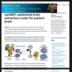
The script has now been tested on a set of more than 70 patient brains, and it performs better than all available tools (see figure below). Lutkenhoff ES, Rosenberg M, Chiang J, Zhang K, Pickard JD, Owen AM, Monti MM. (2014) Optimized Brain Extraction for Pathological Brains (optiBET). PLoS ONE 9(12): e115551. doi:10.1371/journal.pone.0115551. Unite de Neuroimagerie Fonctionnelle. Quantitative MRI & voxel-based quantification (VBQ) Anatomical MR imaging has not only become a cornerstone in clinical diagnosis but also in neuroscience research.
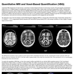
The great majority of anatomical studies rely on T1-weighted images for morphometric analysis of local gray matter volume using voxel-based morphometry (VBM). VBM provides insight into macroscopic volume changes that may highlight differences between groups; be associated with pathology or be indicative of plasticity. A complimentary approach that has sensitivity to tissue microstructure is high resolution quantitative imaging. Whereas in T1-weighted images the signal intensity is in arbitrary units and cannot be compared across sites or even scanning sessions, quantitative imaging can provide neuroimaging biomarkers for myelination, water and iron levels that are absolute measures comparable across imaging sites and time points.
To analyse the quantitative MPM data, we have also created a toolbox specifically for use with the MPM acquisition protocol. Primary contact. MIALab. Available Software: Group ICA Toolbox (Includes GIFT andEEGIFT): Group ICA Toolbox is a MATLAB toolbox which implements multiple algorithms for independent component analysis and blind source separation of group (and single subject) functional magnetic resonance imaging data and electro encephalogram data.
Group ICA Toolbox is listed on NITRC . Ischemic and non-ischemic cardiomyopathy. Mahrholdt H, Wagner A, Judd RM, Sechtem U, Kim RJ.
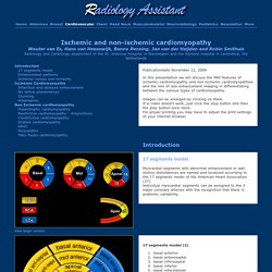
Delayed enhancement cardiovascular magnetic resonance assessment of non-ischaemic cardiomyopathies. Eur Heart J 2005; 26:1461-1474 Delayed Enhancement MR Imaging: Utility in Myocardial Assessment Vogel-Claussen J, Rochitte CE, Wu KC, Kamel IR, Foo TK, Lima JA, Bluemke DA. et al Radiographics 2006; 26:795-810 White JA, Patel MR. The role of cardiovascular MRI in heart failure and the cardiomyopathies. Magn Reson Imaging Clin N Am 2007; 15:541-564 Harris SR, Glockner J, Misselt AJ, Syed IS, Araoz PA.
Cardiac MR imaging of nonischemic cardiomyopathies. M-FFE FE MRI sequence physics and image appearance. Multiple Echo Data Image Combination (MEDIC) is a T2* weighted spoiled gradient echo sequence.
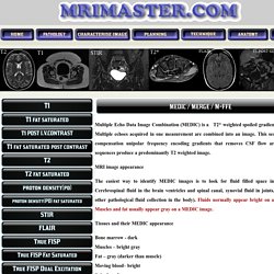
Multiple echoes acquired in one measurement are combined into an image. This sequence uses flow compensation unipolar frequency encoding gradients that removes CSF flow artefacts. MEDIC sequences produce a predominantly T2 weighted image. MRI image appearance The easiest way to identify MEDIC images is to look for fluid filled space in the body (e.g. GDCM: Tool to convert DICOM to DICOM. Gdcmconv [options] file-in file-out The gdcmconv command line program takes as input a DICOM file (file-in) and process it to generate an output DICOM file (file-out). The command line option dictate the type of operation(s) gdcmconv will use to generate the output file. DeveloperWorks : Download: WebSphere Application Server for Developers V8.5. IBM® WebSphere® Application Server Developer Tools provides plug-ins from the Eclipse Marketplace that can be installed into an existing Eclipse environment to support development for WebSphere Application Server.
This no-charge offering compliments the IBM WebSphere Application Server for Developers to provide a lightweight, development environment for the developer desktop. WebSphere Application Server for Developers includes a Liberty Profile that optimizes developer productivity and web application deployment with the new Liberty Profile option, an ultra lightweight, fast starting, highly composable application server profile. The Liberty profile can be downloaded separately or with WebSphere Application Server for Developers. IBM WebSphere Application Server for Developers is a no-charge WebSphere Application Server development runtime for projects that don't warrant the expense of a priced and supported runtime on the developer desktop.
Product specs. Our Solutions - Enlitic. Synchropet. MRI pathology images. Brian Hargreaves - MRI Tools. Www.mytrialwire.com - clever DICOM tool. Welcome - The MRI Blog Network. HONConduct419548 - MRI Blog - HONcode certificate: respect of the 8 HONcode principles by the health website. MRI Mobile. Revising MRI. Fonts, like all of us, have a particular character.
Pun intended. Their shapes can suggest a tone of voice, encourage a level of formality or urgency, or trigger associations with other products or brands. Some allow for fast reading on screens, or easier prolonged reading on paper. Stan's NMR Blog. A personal view of the state of NMR at the start of 2015 NMR is presently going through an unusual phase. For a number of reasons, and for the first time in history, traditional NMR spectroscopy is commercially unprofitable. It was totally abandoned by one major manufacturer, while others are anything but enthusiastic about it. At the same time, there are many new commercial ventures aimed at considerably different types of NMR instruments (compact, sub-compact, table-top, cryogen-free, portable, ex situ, ffc, ...). MRI BLOG: Download Page. Sunday, August 30, 2009 Download Page. Radiology MRI. PractiCal fMRI: the nuts & bolts: GRAPPA.
Info on ferromagnetic detection and MRI safety & screening. Medical Resources and Information. Imaging Analytics University. Zebra Medical Vision. Focus on what’s important – pure research. No need to spend months or years gathering data, building and maintaining an IT and computing infrastructure or worrying about storage costs. Our platform provides access to millions of anonymized, indexed patient imaging studies, reports and associated clinical information. It is highly flexible, allowing development in all major computer vision and machine learning tools, fully web based and fosters collaboration, with full security and privacy features. Academic Institutions and Researchers. Enlitic Premier Program - Enlitic. What is the Premier Partner Program? The Premier Partner Program provides early access to Enlitic's deep-learning based technologies.
MRI-TECH Canada Inc. (MTC) Home.