
Cyclic AMP inhibitors inhibits PDGF-stim... [Cell Signal. 1997 May-Jun. cAMP-dependent pathway. In the field of molecular biology, the cAMP-dependent pathway, also known as the adenylyl cyclase pathway, is a G protein-coupled receptor-triggered signaling cascade used in cell communication.[1] Mechanism[edit] G protein-coupled receptors (GPCRs) are a large family of integral membrane proteins that respond to a variety of extracellular stimuli.
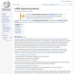
Each GPCR binds to and is activated by a specific ligand stimulus that ranges in size from small molecule catecholamines, lipids, or neurotransmitters to large protein hormones. Cyclic adenosine monophosphate. Cyclic adenosine monophosphate (cAMP, cyclic AMP or 3'-5'-cyclic adenosine monophosphate) is a second messenger important in many biological processes. cAMP is derived from adenosine triphosphate (ATP) and used for intracellular signal transduction in many different organisms, conveying the cAMP-dependent pathway.
History[edit] Earl Sutherland of Case Western Reserve University won a Nobel Prize in Physiology or Medicine in 1971 "for his discoveries concerning the mechanisms of the action of hormones," especially epinephrine, via second messengers (such as cyclic adenosine monophosphate, cyclic AMP). Synthesis and decomposition[edit] cAMP decomposition into AMP is catalyzed by the enzyme phosphodiesterase. Functions[edit] Role of cAMP in eukaryotic cells[edit] cAMP and its associated kinases function in several biochemical processes, including the regulation of glycogen, sugar, and lipid metabolism. Regulation of Adenylyl Cyclases.
The animation below depicts the regulation of adenylyl cyclase by hormone stimulation. 1.
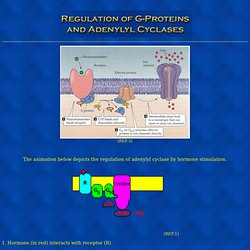
Hormone (in red) interacts with receptor (R) 2. Receptor (R) binds to G complex ( Gb(beta & gamma)-Ga(alpha)) 3. 4. 5. 6. The images below show the regulation of an adenylate cyclase in a step by step manner: Step #1: Resting state- receptor binding site is unoccupied. Step #2: Bound ligand. Step #3: GTP/GDP exchange and adenylate cyclase activation. Structural Biochemistry/Cell Signaling Pathways/Endocrine Signaling.
The word endocrine is actually from the Greeks and the endo definition means "within" and krine definition means "to separate or to secrete.
" The term endocrine means “secreting internally,” and specifically refers to secretions that are distributed in the body by way of the bloodstream. Endocrine cells are made up of ductless glands that produce chemical messages called hormones, which are released into the internal environment of the body. These Endocrine secretions are distinguished from exocrine secretions, which are released to the external environment. Thus, endocrine signaling occurs when endocrine cells release hormones that act on distant target cells in the body. Structural Biochemistry/Cell Signaling Pathways/Endocrine Signaling. G protein coupled receptor. The seven-transmembrane α-helix structure of a G protein-coupled receptor There are two principal signal transduction pathways involving the G protein-coupled receptors: the cAMP signal pathway and the phosphatidylinositol signal pathway.[6] When a ligand binds to the GPCR it causes a conformational change in the GPCR, which allows it to act as a guanine nucleotide exchange factor (GEF).
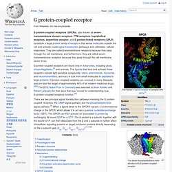
The GPCR can then activate an associated G-protein by exchanging its bound GDP for a GTP. The G-protein's α subunit, together with the bound GTP, can then dissociate from the β and γ subunits to further affect intracellular signaling proteins or target functional proteins directly depending on the α subunit type (Gαs, Gαi/o, Gαq/11, Gα12/13).[7]:1160 Classification[edit] Classification Scheme of GPCRs. In all, GPCRs can be grouped into 6 classes based on sequence homology and functional similarity:[9][10][11][12]
Sturcture/form/function. As their name implies, GPCRs interact with G proteins in the plasma membrane.

When an external signaling molecule binds to a GPCR, it causes a conformational change in the GPCR. This change then triggers the interaction between the GPCR and a nearby G protein. G proteins are specialized proteins with the ability to bind the nucleotides guanosine triphosphate (GTP) and guanosine diphosphate (GDP). Some G proteins, such as the signaling protein Ras, are small proteins with a single subunit. However, the G proteins that associate with GPCRs are heterotrimeric, meaning they have three different subunits: an alpha subunit, a beta subunit, and a gamma subunit. Steroid hormone receptor. Protein Hormone Receptor. A hormone receptor is a molecule that can bind to a specific hormone.
Receptors for peptide hormones tend to be found on the plasma membrane of cells, whereas receptors for lipid-soluble hormones are usually found within the cytoplasm. Upon hormone binding, the receptor can initiate multiple signaling pathways which ultimately lead to changes in the behaviour of the target cells. Water-soluble hormone receptors[edit] Water-soluble hormones include glycoproteins, catecholamines and peptide hormones composed of polypeptides, e.g. thyroid-stimulating hormone, follicle-stimulating hormone, leutinizing hormone and insulin.
These molecules are not lipid-soluble and therefore cannot diffuse through cell membranes. What is a steroid. Photo by: johnnychaos Steroids are a family of lipid molecules that includes cholesterol, steroid hormones, and bile salts.
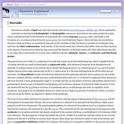
These amphipathic molecules (containing both hydrophobic and hydrophilic regions) are derived from two-carbon acetyl-CoA units, whose combination leads to the formation of isoprenoids (five-carbon isoprene molecular units), and finally to the formation of a seventeen-carbon tetracyclic hydrocarbon, the steroid skeleton. Figure 1 shows the basic steroid skeleton structure, made up of three six-membered rings and one five-membered ring. Inositol trisphosphate. The inositol trisphosphate trianion Properties of IP3 Chemical formula and molecular weight IP3 is a polyatomic ion with a molecular mass of 420.10 g/mol.
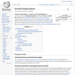
Its empirical formula is C6H15O15P3. It is composed of an inositol ring with three phosphate groups bound at the 1, 4, and 5 carbon positions, and three hydroxyl groups bound at positions 2, 3, and 6.[1] Chemical proprieties of phosphate group and IP3 Phosphate groups can exist in three different forms depending on a solution's pH.