
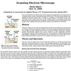
Professor Robert B. Laughlin, Department of Physics, Stanford University. Sheila Bijoor May 11, 2008 (Submitted as coursework for Applied Physics 273, Stanford University, Spring 2007)
Untitled Document. A paper presented at the Annual Conference of the Electron Microscope Group of the Institute of Physics: EMAG '97 (For full details see Conference Proceedings).
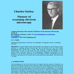
Copyright © 1997 IOP Publishing Ltd K C A Smith Engineering Department, University of Cambridge, Trumpington Street, Cambridge CB2 1PZ ABSTRACT: Charles Oatley stands with Manfred von Ardenne as one of the two great pioneers of scanning electron microscopy. His involvement with the SEM began immediately after World War II when, fresh from his wartime experience in the development of radar, he perceived that new techniques could be brought to bear which would overcome some of the fundamental problems encountered by von Ardenne in his pre-war research on the instrument. History of electron microscopy, 1931-2000. More detailed story here, summary below. 1.
Microscopy Books. The History of Electron Microscope. A brief history of Micorscopy - Biological Electron Microscopy. Microscopy and Microanalysis - Abstract - Key Events in the History of Electron Microscopy. Energy filter. The History of Scanning Electron Microscopes" The development of SEMs started with more of a whimper than a bang.
When the technology was first unveiled in 1935, a group of marketing professionals was asked to evaluate the new instrument's potential in the marketplace. After polling the scientific community, the marketing experts weren't too optimistic. They estimated a need for, at most, 10 of the devices worldwide. As it turns out, the experts vastly underestimated the potential of SEMs, and thankfully, their dour outlook failed to deter further development of the technology. As a result, more than 50,000 SEMs fill laboratories and businesses across the globe [source: Breton]. For one thing, scientists had pushed optical microscopes to their limits. Since development of TEMs was well under way by the time SEMs came along, the latter were initially considered unnecessary. History of electron microscopy, 1931-2000. History of electron microscopy.
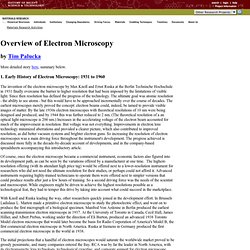
Most of the information in the above table was taken from G.
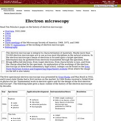
Thomas, "The impact of electron microscopy on materials research", in Impact of Electron and Scanning Probe Microscopy on Materials Research, edited by Rickerby, Valdre, and Valdre, Kluwer: 1999; and from Vander Voort & Friel (eds.), Development in Materials Characterization Technologies. The image on the right is also taken from the latter (Figure 1, page 45) by permission of ASM International, Materials Park, OH 44073-0002. The use of the tree as a metaphor for the historical development is interesting. It is certainly suggestive, and as such it does not matter greatly whether the metaphor applies to all facets.
What is interesting about the history of electron microscopy? German engineer Max Knoll and physicist Ernst Ruska are recognized for the development of the first electron microscope, in 1932, with Ruska being awarded the Nobel Prize in 1986 for his work in electron optics.
Several significant historical events and thought processes led to the development of the electron microscope: • In 1873, physicists Hermann von Helmholtz and Ernst Abbe demonstrate that optical resolution depends on the wavelength of the illumination source (see also: Fig. 2, in "How is EM different from light microscopy? "). This insight leads to the what-if notion of an electron microscope (“What if you could use illumination of wavelength that is smaller than light?”).
However, even though Helmholtz argues for “atoms of electricity” in 1881, the existence of electrons is not demonstrated until experiments carried out by J.J. Thomson and colleages in 1896. Figure 2. Electron microscope. Diagram of a transmission electron microscope An electron microscope (EM) is a type of microscope that uses an electron beam to illuminate a specimen and produce a magnified image.
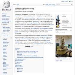
An EM has greater resolving power than a light microscope and can reveal the structure of smaller objects because electrons have wavelengths about 100,000 times shorter than visible light photons. They can achieve better than 50 pm resolution[1] and magnifications of up to about 10,000,000x whereas ordinary, non-confocal light microscopes are limited by diffraction to about 200 nm resolution and useful magnifications below 2000x. The electron microscope uses electrostatic and electromagnetic lenses to control the electron beam and focus it to form an image.
These electron optical lenses are analogous to the glass lenses of a light optical microscope. History[edit] Dennis McMullan Scanning Microscope. Full version of a presentation at the 51st Annual Meeting of the Microscopy Society of America, Cincinnati, August 1993.
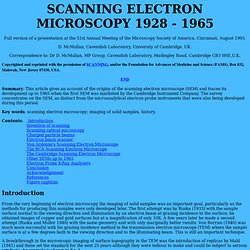
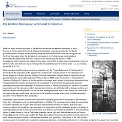
D. McMullan, Cavendish Laboratory, University of Cambridge, UK. Correspondence to: Dr D. RCA EMU-2B transmission electron microscope, United States, 1946. The Electron Microscope, A Personal Recollection — Department of Physics. By H.L.Watson Introduction Fig. 1a: The 1938 Toronto model.
Laboratorio de Microscopía de Alta Resolución. It is curious than, however, these German pioneers did not know nothing at all about Louis de Broglie's theory before they had constructed their first electron microscope.
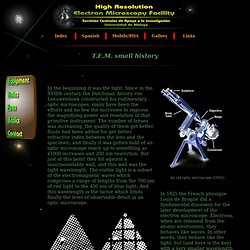
In fact, for discretion, they avoided using this term when exposing his work, although at a later time this was considered like the first electron microscope of history (very at a later time, since Ruska had to expect 55 years until he won the Nobel Prize in 1986). A year after this first prototype they wrote on paper the concept of electron microscope, and in only one more year (1933) they constructed an electron microscope of three lenses which gets 12000 increases and a resolution of 50nm.
They was going beyond the optic microscopy. Since then, several companies began to manufacture and to distribute the first commercial electron microscopes: Metropolitan Vickers ( 1936 ), Siemens ( 1939 ), RCA ( 1941 ), Hitachi ( 1941 ), Philips ( 1949 ), JEOL ( 1949 ), etc. Laboratorio de Microscopía de Alta Resolución.