
UConn unveils ambitious knee regeneration research. The University of Connecticut announced the launch of its new grand research challenge: regeneration of a human knee within 7 years, and an entire limb within 15 years.

The major international research undertaking is called The HEAL Project: Hartford Engineering a Limb. According to UConn, it is the brainchild of UConn Health's Cato T. Laurencin, a leading surgeon-scientist in orthopaedic surgery, engineering, and the new field of regenerative engineering. His laboratory research successes include the growth of bone and knee ligaments. For the project, Laurencin is teaming with other tissue engineering, regenerative medicine, and bioengineering experts dedicated to the mission of advancing the fields and developing future therapies for patients living with musculoskeletal defects or who have limb injury or loss. "The launch of the HEAL Project is a transformative moment for science and medicine," said Laurencin.
Read more Quick helps Frontier hold steady in CT. League of Denial: The NFL's Concussion Crisis. Microtubule. BC Online: 9E - Memory and Learning in Aplysis. This page has been compiled from several web pages and is not meant to be a cohesive chapter.
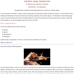
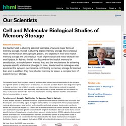
For our last class we will listen to a tape of a talk, Remembrance of Things Past, by Nobel Laureate Eric Kandel. He won the 2000 prize in Medicine and Physiology for his study of memory and learning, especially in the sea snail, Aplysia. The information in the following page comes from web links (references) and are threaded together to present the background to the talk we will hear on Monday. Please read this thoroughly so you can be prepared to get the most out of his talk. Eric R. Kandel, MD Research Abstract. The general finding that long-term plasticity and long-term memory recruit transcription in the nucleus, an organelle shared by all synapses of a neuron, has raised a question that we have begun to explore in Aplysia and mice: Are long-term changes cell-wide, or can induced gene products be spatially compartmentalized so that they selectively alter the function of some synapses and not others?
In mice, we have also explored the molecular mechanisms whereby attention modifies and stabilizes internal representation of space. Gap junction - Wikipedia. A gap junction may also be called a nexus or macula communicans.
When found in neurons or nerves it may also be called an electrical synapse. While an ephapse has some similarities to a gap junction, by modern definition the two are different. One gap junction channel is composed of two connexons (or hemichannels), which connect across the intercellular space.[4][5][6] Gap junctions are analogous to the plasmodesmata that join plant cells.[7] One of the young boys in my family is currently all about dinosaurs (and monster trucks, living up to all little boy traditions).

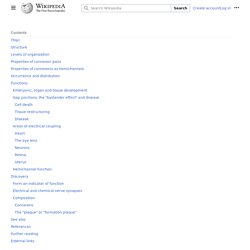
He is knowledgable enough about them to name the lovely specimen with the incredibly long neck an Apatosaurus, not Brontosaurus, putting his knowledge a caliber above mine at that age. But he may yet be indoctrinated with another of the great dinosaur myths, that each stegosaurus has a second brain in its tail, because even “scientific” documentaries featuring respected researchers get twisted to perpetuate that idea. Perhaps those dinosaur documentary makers are just jealous that the humble octopus turns out to be so much cooler than their great lizards. Your average octopus might not seem like a cognitive juggernaut, especially not in an ocean that also contains dolphins, our favorite frontrunner for animal intelligence.
OCTOPUSES & RELATIVES: THE "INTELLIGENT" OCTOPUS. Octopuses have long been thought to be incapable of using their arms and suckers for manipulative tasks, such as removing a cork stopper from a glass jar to capture a crab inside.
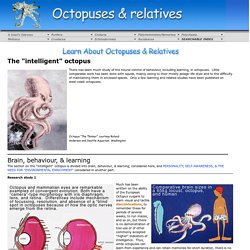
That they occasionally do so was credited to happenstance. At least part of the reason for this is that octopuses seem to lack the proprioceptive ability to assess the positions of their arms in space. So, removing a screw-top lid from a jar to capture a crustacean inside would not be possible…or would it? Perhaps the appropriate stimuli have never been presented. Cephalove: A View of the Octopus Brain. In this post, I am going to outline octopus neuroanatomy, to the best of my ability.

It's a complicated subject that I am only beginning to have a grasp on, but I want to post more about specific research papers regarding cephalopod brains, so I figure I should review this first. Let's get right to it. This figure is from J. Z. Opsin/vitamin A based photopigments in extraretinal photoreceptors — Nuffield Department of Clinical Neurosciences. Non-mammalian vertebrates possess a diverse complement of photoreceptors including the pineal organ, deep encephalic photoreceptors, and dermal/peripheral tissue photoreceptors.

Although these photoreceptors were first recognised in the early part of the 19th century, the basis for their photosensitivity remained (and to a degree still remains) poorly understood. My work provided overwhelming evidence that these diverse photoreceptors use broadly conserved mechanisms based upon opsin/vitamin A photopigments. Two of my early Nature papers, summarise these conceptual breakthroughs:
Louis J. Guillette, Jr., Ph.D. - Featured Expert - MUSC. Oligodendrocytes - structure/function. Spinal nerve connections. Cranial nerves anatomy Page 70. Virtual Lab: Tyrannosaurus Rex Brain. Recommended Reading Scientists create detailed map of dinosaur brain, Science, theguardian.com Witmer Lab, Perspectives Advanced Reading Witmer and Ridgely, Anatomical Record: Description of T.
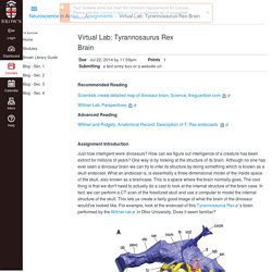
Rex endocasts. Assignment Introduction Just how intelligent were dinosaurs? Endocast of Tyrannosaur brain. Source: Witmer and Ridgely, 2009, The Anatomical Record. Superior colliculus - Wikipedia. The superior colliculus, (Latin, upper hill) is a paired structure of the mammalian midbrain.
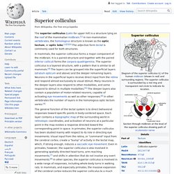
In other vertebrates the homologous structure is known as the optic tectum or simply tectum. The adjective form tectal is commonly used for mammals as well as other vertebrates. Opt 662 #15 Retino-Tectal Pathway 2015. Superior colliculus - Wikipedia. Superior colliculus - Wikipedia. 1302 Brain Vesicle DevN - Neuraxis - Wikipedia. The Blood Supply of the Brain and Spinal Cord - Neuroscience - NCBI Bookshelf. Cerebral cortex - Wikipedia. Outer layer of the cerebrum of the mammalian brain. Cerebral cortex - Wikipedia. Paleoencephalon - Wikipedia. In the anatomy of animals, paleoencephalon refers to most regions in the brain that are not part of the neocortex or neoencephalon. The paleoencephalon is the phylogenetically oldest part of the animal brain.
Paleoenchepheal areas of older species are larger in proportion to overall brain volume as compared to those of mammals. Allocortex - Wikipedia. The specific regions of the brain usually described as belonging to the allocortex are the olfactory system, and the hippocampus. Allocortex is termed heterogenetic cortex, because during development it never has the six-layered architecture of homogenetic neocortex. It differs from heterotypic cortex, a type of cerebral cortex, which during prenatal development, passes through a six-layered stage to have fewer layers, such as in Brodmann area 4 that lacks granule cells.[2] Structure[edit] The allocortex has just three or four layers of neuronal cell bodies in contrast to the six layers of the neocortex.
There are three subtypes of allocortex, the paleocortex, archicortex and periallocortex.[3] Hippocampus - Wikipedia. The hippocampus (from the Greek ἱππόκαμπος, "seahorse") is a major component of the brain of humans and other vertebrates. Humans and other mammals have two hippocampi, one in each side of the brain. The hippocampus is part of the limbic system, and plays important roles in the consolidation of information from short-term memory to long-term memory, and in spatial memory that enables navigation. The hippocampus is located under the cerebral cortex in the allocortex,[1][2][3] and in primates it is in the medial temporal lobe. It contains two main interlocking parts: the hippocampus proper (also called Ammon's horn)[4] and the dentate gyrus. Since different neuronal cell types are neatly organized into layers in the hippocampus, it has frequently been used as a model system for studying neurophysiology. Spinal Cord Injury Levels & Classification.
Wise Young, Ph.D., M.D.W. M. Keck Center for Collaborative Neuroscience Rutgers University, Piscataway, NJ People with spinal cord injury are often told that they have an injury at a given spinal cord level. Fibroblast growth factors and their receptors in the central nervous system. Nervous Tissue - Structure and Functions of Human Tissue Types. Note: This page is part of the section about the structure and function of different Tissue Types, which is related to the section about Histology and Cells (incl. structure of animal cells, cell division, mitosis, meiosis). Meninges. Astrocyte - Wikipedia. Research since the mid-1990s has shown that astrocytes propagate intercellular Ca2+ waves over long distances in response to stimulation, and, similar to neurons, release transmitters (called gliotransmitters) in a Ca2+-dependent manner. Gap-43 protein - Wikipedia. Growth Associated Protein 43 also known as GAP43 is a protein that in humans is encoded by the GAP43 gene.[3] GAP43 has been termed a 'growth' or 'plasticity' protein because it is expressed at high levels in neuronal growth cones during development, during axonal regeneration and is phosphorylated after long-term potentiation (LTP) and after learning (reference needed).
This protein is considered a crucial component of the axon and presynaptic terminal, its null mutation leading to death within days after birth due to axon pathfinding defects.[4] Synonyms[edit] Glioma Cell - Cerca con Google. A canine BCAN microdeletion associated with episodic falling syndrome - UCL Discovery. Gill, JL; Tsai, KL; Krey, C; Noorai, RE; Vanbellinghen, JF; Garosi, LS; Shelton, GD; Gill, JL; Tsai, KL; Krey, C; Noorai, RE; Vanbellinghen, JF; Garosi, LS; Shelton, GD; Clark, LA; Harvey, RJ; - view fewer (2012) A canine BCAN microdeletion associated with episodic falling syndrome. Keratan sulfate. Structure[edit] Hearing and auditory system: morawski_pictures. Hearing and auditory system: Markus Morawski. About 20% of central nervous system (CNS) volume is extracellular matrix (ECM) which is composed of specific macromolecules filling the extracellular space and being considered as an essential determinant of the organization and function of the CNS.
Glycosaminoglycan. Chondroitin sulfate. Chemical structure of one unit in a chondroitin sulfate chain. Porifera - Nervous System. Sea sponges have the makings of a nervous system. Megakaryocyte. Résultats Google Recherche d'images correspondant à. The Shoichet Lab. Astrogliosis. Human Antatomy & Physiology Nervous Tissue Chapter 12 By Abdul Fellah, Ph.D Copyright (c) The McGraw-Hill Companies, Inc. Permission required for reproduction. - ppt download. [Biogenesis of melanosomes - the chessboard of pigmentation]. Nanoparticles for Neurotherapeutic Drug Delivery in Neurodegenerative Disorders - Brain Neurotrauma - NCBI Bookshelf. Patent US9259419 - Compositions and methods relating to solenopsins and their uses in treating ... - Google Patents.
Making the Impossible Probable. Chapter 143. The Spinal Cord: Neural Crest Cells and Myelination - Review of Medical Embryology Book - LifeMap Discovery. THE BRAIN FROM TOP TO BOTTOM. File:1801 The Endocrine System.jpg. Hepatic Nerves - Hepatic Circulation - NCBI Bookshelf. Spinal Cord - Embryonic Development & Stem Cells - LifeMap Discovery. Neural Crest - Embryonic Development & Stem Cells - LifeMap Discovery. Choroid plexus.