
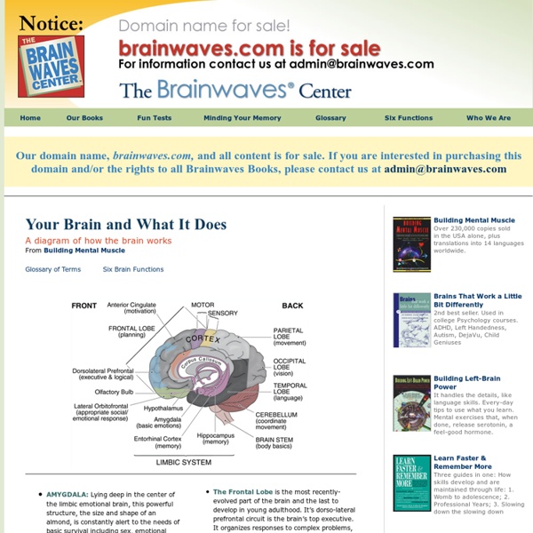
Diagram of Brain In this diagram of the brain the different sections are shown. The Cerebrum are the two large hemispheres of the brain. Each hemisphere is further divided into lobes. Above is the break up of where each lobe is located and the structures under the cerebrum that make up the rest of the brain. Diagramed above are some of the major cortexes and their locations. This is what's known as a sagittal view of the brain. We recommend investing in this guide to human physiology and anatomy. We recommend investing in this guide to human physiology and anatomy.
Brain Gym® Exercises By Kenneth Beare Updated December 16, 2014. Brain Gym® exercises are exercises designed to help the brain function better during the learning process. As such, you can think of Brain Gym® exercises as part of the overall theory of multiple intelligences. These exercises are based on the idea that simple physical exercise helps blood flow to the brain and can help improve the learning process by making sure the brain stays alert. Students can use these simple exercises on their own, and teachers can use them in class to help keep energy levels up throughout the day. These simple exercises are based on the copyrighted work of Paul E. continue reading below our video Loaded: 0% Progress: 0% Below is a series of movements called PACE. Drink Water As Carla Hannaford says, "Water comprises more of the brain (with estimates of 90%) than of any other organ of the body." Drinking water is very important before any stressful situation - tests! Brain Buttons Cross Crawl Stand or sit. Hook Ups Here
CogniFit Brain Fitness And Memory Programs, Brain Training Brain Functions and Diagram Brain Functions and Diagram Recognizing where damage is located and understanding the effects Neuro Point of View by: Dr. William Winslade Among the most devastating effects of head injury is the secondary damage that can follow hours and days later. Scientists still don't understand how the brain heals itself, so long-term recovery from any traumatic brain injury is uncertain, and its course is difficult to predict. Recovery from traumatic brain injury may be quick or slow; it may be complete, partial, or absent. People who do recover from traumatic brain injury must be highly motivated and persistent. The most comforting stories about recovery from traumatic brain injury have the timeless power of great myths. Dr.
Home - Gymnasium for Brain Brain Games for Adults Brain games are fun to play at gatherings, meetings, kitty parties or on outings. Most of the brain games usually include trick questions and brain teasers, or some very hard riddles. The following are some brain games that you can play in both small and large groups. These games test some of the capabilities of the brain like concentration, memorization and flexibility. The Stroop Test The Stroop test is a brilliant way to test a person's mental flexibility and concentration. Analogy This game is a favorite among kids, but can be played by adults too. The Face of the Penny This is a very nice brain game for adults. Concentrating on Numbers Among all the brain games for adults, this one is rather popular. If you are in a small group then it would be appropriate to play some nice math riddles and puzzles.
Fear Can Be Erased from the Brain Newly formed emotional memories can be erased from the human brain. This is shown by researchers from Uppsala University in a new study now being published by the academic journal Science. The findings may represent a breakthrough in research on memory and fear. Thomas Ågren, a doctoral candidate at the Department of Psychology under the supervision of Professors Mats Fredrikson and Tomas Furmark, has shown, that it is possible to erase newly formed emotional memories from the human brain. When a person learns something, a lasting long-term memory is created with the aid of a process of consolidation, which is based on the formation of proteins. In the study the researchers showed subjects a neutral picture and simultaneously administered an electric shock. In the study the researchers showed subjects a neutral picture and simultaneously administered an electric shock. ‘These findings may be a breakthrough in research on memory and fear. Notes about this fear and anxiety research
BrainBuilder BrainBuilder® is the brain fitness program designed to improve your memory, attention and brainspeed! For children, teens and adults! Experience these REMARKABLE RESULTS with BrainBuilder: Better memory and attentionSharper focus and clearer thinkingImproved visual and auditory processingIncreased BrainSpeedStronger problem-solving skills Brain Coach The Coach will test your BrainSpeed, design your daily sessions, track your improvement, adjust your training program and motivate you along the way. Brain Exercises Train your brain with a library of more than 20 diverse, visual, auditory and focus exercises. Brain Music Each session begins and concludes with listening to psychoacoustically-based classical music produced with visual imagery to relax your body and alert your mind for optimal cognitive performance. Brain Games Enjoy fun and interactive brain games – each designed to engage and challenge you. Practice Area Progress Charts Results are measurable! Keep Your Brain Performing at Its Best! 1.
Cognitive Rehabilitation Word Game Quizzes Cognitive rehabilitation can be helped by performing regular cognitive exercises that force the brain to work a little. This simple word game quiz provides just such a workout, but can be done in the non-threatening environment of your home. Since you can download and print the quizzes, they can be used over and over again. Each quiz contains a tracking line if you want to use it to measure progress. For instance, you can take Quiz One as many times as you wish. Some of the quizzes are more difficult than others, but they all involve finding words or characters and drawing a circle around them. Before you jump down to look at the solution, just take a moment or two to see which words you can find. Download Quizzes for Your Use To use these quizzes, all you need is a computer, a pdf reader and a printer. Click here to download Cognitive Rehabilitation Quiz One. Click here to download Cognitive Rehabilitation Quiz Ten. Click here to download Cognitive Rehabilitation Quiz Twenty.
Short-term memory is based on synchronized brain oscillations Scientists have now discovered how different brain regions cooperate during short-term memory. Holding information within one's memory for a short while is a seemingly simple and everyday task. We use our short-term memory when remembering a new telephone number if there is nothing to write at hand, or to find the beautiful dress inside the store that we were just admiring in the shopping window. Yet, despite the apparent simplicity of these actions, short-term memory is a complex cognitive act that entails the participation of multiple brain regions. However, whether and how different brain regions cooperate during memory has remained elusive. A group of researchers from the Max Planck Institute for Biological Cybernetics in Tübingen, Germany has now come closer to answering this question. It has long been known that brain regions in the frontal part of the brain are involved in short-term memory, while processing of visual information occurs primarily at the back of the brain.
brainsource.com | Exploring the Human Brain Free Brain Games Training Online - Improve Memory, Have Fun! Training your brain with free online brain games is a fun way to keep your mind active and potentially improve your memory, concentration, and other brain skills. There are now over 250 free brain training games on this site. Not sure where to start? Check out the most popular games. Also see the game categories in the sidebar at right and in the menu above. Examples of popular games include Scrabble Sprint, Butterfly Connect, and Basic Solitaire. To play these online games, an up-to-date version of the free Adobe Flash Player browser plug-in must be installed in your browser. If the games won't open for you, there might be an issue with your browser. If you still have problems accessing the games, check out my troubleshooting page or feel free to contact me directly for assistance. You can start your own brain training program right now. To keep your mind in top shape, play brain games often. For a full-brain workout, play a variety of games. Which Brain Skills Can You Improve? Prof.
Schizophrenia diagnosis associated with progressive brain changes among adolescents Adolescents diagnosed with schizophrenia and other psychoses appear to show greater decreases in gray matter volume and increases in cerebrospinal fluid in the frontal lobe compared to healthy adolescents without a diagnosis of psychosis, according to a report in the January issue of Archives of General Psychiatry, one of the JAMA/Archives journals. “Progressive loss of brain gray matter (GM) has been reported in childhood-onset schizophrenia; however, it is uncertain whether these changes are shared by pediatric patients with different psychoses,” the authors write as background information in the study. Celso Arango, M.D., Ph.D., of the Hospital General Universitario Gregorio Marañón, Madrid, Spain, and colleagues, examined the progression of brain changes in first-episode early-onset psychosis and the relationship to diagnosis and prognosis at two-year follow-up among patients at six child and adolescent psychiatric units in Spain.
Brain Training | Brain Exercises | Brain Fitness Games | Brain Metrix