
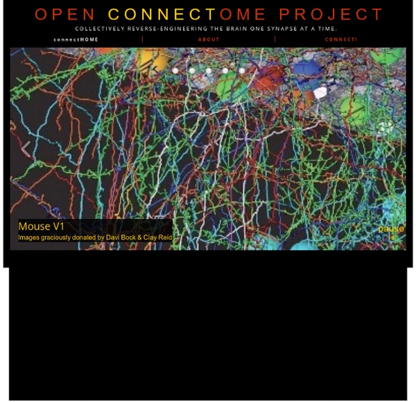
The Connectome — Harvard School of Engineering and Applied Sciences Lead investigators Hanspeter Pfister (SEAS ), Jeff Lichtman (FAS/Molecular & Cellular Biology, Center for Brain Science) and Clay Reid (HMS/Neurobiology, Center for Brain Science) Description The overall goal of the Connectome project is to map, store, analyze and visualize the actual neural circuitry of the peripheral and central nervous systems in experimental organisms, based on a very large number of images from high-resolution microscopy.
Neur-one Connectomics Connectomics is the production and study of connectomes: comprehensive maps of connections within an organism's nervous system, typically its brain or eye. Because these structures are extremely complex, methods within this field use a high-throughput application of neural imaging and histological techniques in order to increase the speed, efficiency, and resolution of maps of the multitude of neural connections in a nervous system. While the principal focus of such a project is the brain, any neural connections could theoretically be mapped by connectomics, including, for example, neuromuscular junctions. Tools[edit] Model Systems[edit] Aside from the human brain, some of the model systems used for connectomics research are the mouse,[3] the fruit fly,[4] the nematode C. elegans,[5][6] and the barn owl.[7] Applications[edit] Criticism[edit] Comparison to genomics[edit] See also[edit] References[edit] Further reading[edit] External links[edit]
Neuranat Morphologie Ce chapitre aborde les bases de l'anatomie descriptive du crâne, des méninges et du système nerveux central. Atlas Ce chapitre propose des atlas anatomiques interactifs dans les trois plans, un atlas neuroradiologique avec curseur 3D synchronisé dans les trois plans de l'espace et un atlas du tronc cérébral de l'espace neuroradiologiques, montrant un point choisi au niveau du cerveau simultanément dans les 3 plans de l'espace et sur une reconstruction 3D ainsi qu'un atlas du tronc cérébral. Vidéos Le chapitre Vidéos constitue un véritable atelier de dissection du cerveau grâce à des documents richmédia associant simultanément vidéo, images et textes. Animations Retrouvez des animations concernant la fissure choroïdienne, le fornix et le quatrième ventricule et un "serious game", Dyn@slice, permettant de s'exercer à remettre dans l'ordre des coupes de cerveau. © UPMC.
home IBM scientists create most comprehensive map of the brain’s network "The Mandala of the Mind": The long-distance network of the Macaque monkey brain, spanning the cortex, thalamus, and basal ganglia, showing 6,602 long-distance connections between 383 brain regions. (PNAS) The Proceedings of the National Academy of Sciences (PNAS) published Tuesday a landmark paper entitled “Network architecture of the long-distance pathways in the macaque brain” (an open-access paper) by Dharmendra S. Modha (IBM Almaden) and Raghavendra Singh (IBM Research-India) with major implications for reverse-engineering the brain and developing a network of cognitive-computing chips. “We have successfully uncovered and mapped the most comprehensive long-distance network of the Macaque monkey brain, which is essential for understanding the brain’s behavior, complexity, dynamics and computation,” Dr. Modha says. The center of higher cognition and consciousness? Core subnetwork (PNAS) Prefrontal cortex: integrator-distributor of information Dr. Amara D.
Simple mathematical pattern describes shape of neuron ‘jungle’ Neuron shape model: target points (red) distributed in a spherical volume and connected to optimize wiring in a tree (black) (credit: H. Cuntz et al./PNAS) University College London (UCL) neuroscientists have found that there is a simple pattern that describes the tree-like shape of all neurons. Neurons look remarkably like trees, and connect to other cells with many branches that effectively act like wires in an electrical circuit, carrying impulses that represent sensation, emotion, thought and action. Over 100 years ago, Santiago Ramon y Cajal, the father of modern neuroscience, sought to systematically describe the shapes of neurons, and was convinced that there must be a unifying principle underlying their diversity. Cajal proposed that neurons spread out their branches so as to use as little wiring as possible to reach other cells in the network. New work by UCL neuroscientists has revisited this century-old hypothesis using modern computational methods.
First map of the human brain reveals a simple, grid-like structure between neurons In an astonishing new study, scientists at the National Institutes of Health (NIH), have imaged human and monkey brains and found… well, the image above says it all. It turns out that the pathways in your brain — the connections between neurons — are almost perfectly grid-like. It’s rather weird: If you’ve ever seen a computer ribbon cable — a flat, 2D ribbon of wires stuck together, such as an IDE hard drive cable — the brain is basically just a huge collection of these ribbons, traveling parallel or perpendicular to each other. There are almost zero diagonals, nor single neurons that stray from the neuronal highways. The human brain is just one big grid of neurons — a lot like the streets of Manhattan, minus Broadway, and then projected into three dimensions. This new imagery comes from a souped-up MRI scanner that uses diffusion spectrum imaging to detect the movement of water molecules within axons (the long connections made by neurons). “Before, we had just driving directions.
The Brain CONNECT Project