

Brain Atlas - Introduction The central nervous system (CNS) consists of the brain and the spinal cord, immersed in the cerebrospinal fluid (CSF). Weighing about 3 pounds (1.4 kilograms), the brain consists of three main structures: the cerebrum, the cerebellum and the brainstem. Cerebrum - divided into two hemispheres (left and right), each consists of four lobes (frontal, parietal, occipital and temporal). The outer layer of the brain is known as the cerebral cortex or the ‘grey matter’. It covers the nuclei deep within the cerebral hemisphere e.g. the basal ganglia; the structure called the thalamus, and the ‘white matter’, which consists mostly of myelinated axons. – closely packed neuron cell bodies form the grey matter of the brain. Cerebellum – responsible for psychomotor function, the cerebellum co-ordinates sensory input from the inner ear and the muscles to provide accurate control of position and movement. Basal Ganglia Thalamus and Hypothalamus Ventricles Limbic System Reticular Activating System Neurons Glia
Student Nurse - A&P Anatomy and Sectional Terminology It sounds silly, but you learn as many new words in an A&P course as you do a beginning foreign language course. Really --there's been research to prove it! - Kevin Patton Links here are for general A & P resources. For more detailed system by system info see: Cardiac, Ear, Endocrine, Eye, Gastrointestinal, Hematological, Integumentary, Musculoskeletal, Neurological, Peripheral Vascular, Renal, and Respiratory.
Medicine | Ebooks 2012 German | ISBN: 3709104661 | 2013 | 550 pages | PDF | 11 MB Das Buch liefert eine ubersichtliche und pragnante Darstellung der diagnostischen und therapeutischen Rehabilitationskonzepte fur zahlreiche Krankheitsbilder. Fur die 3. Download [Fast Download] Kompendium Physikalische Medizin und Rehabilitation: Diagnostische und therapeutische Konzepte, Auflage: 3 2012 | 395 Pages | ISBN: 1441908013 | PDF | 11 MB Clinical PET and PET/CT, 2nd Edition presents a valuable overview of the basic principles and clinical applications of PET and PET/CT. Emphasis is placed on the familiarization of normal distribution, artifacts, and common imaging agents such as FDG in conjunction with CT, MRI, and US to establish the clinical effectiveness of PET and PET/CT.
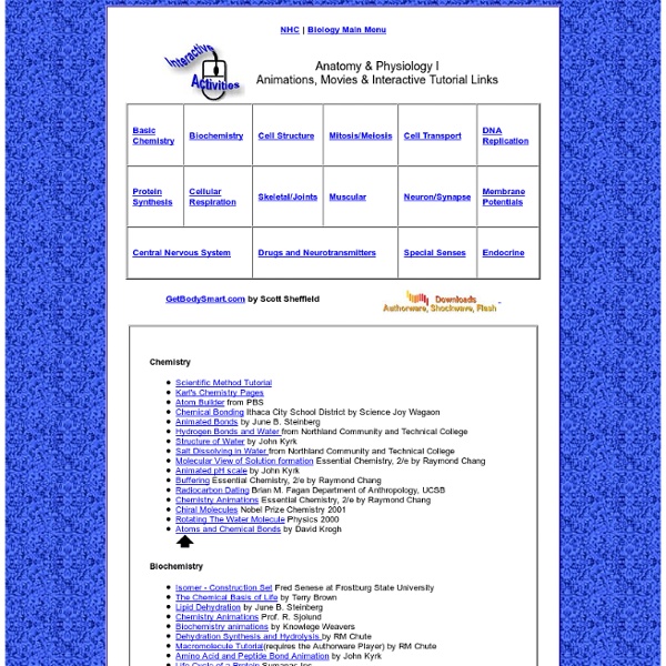
Master Muscle List Home Page IHS - International Headache Society» Home Anatomy and Physiology animations Listed below are a collection of physiology animations and anatomy animations. These animations are intended to support text or lecture and it is important that they are not seen as stand-alone reference material. Notes: If you or your students discover any factual errors in the animations please let me know: andrew@visualization.org.uk Some of the animations can only be accessed from the university network - please contact Liz Hodgson in the LDU if you would like them on WebCT so that students can access them externally. Here are some animations of organs/organ systems: Cranial nerves (No text version) Cranial nerves (Customised version)) Anatomical directions and sections. Central and Peripheral Nervous System Vertebrae: meninges etc. Brain: meninges cerebrospinal fluid etc. Primary motor and somatosensory Cortices (Homunculus) Skin turgor Contraction and electrical activity in the heart (Slowed down representation) Intestinal villi (diagram) Gastrointestinal tract (diagram) Stomach, liver etc.
Anatomy and Physiology Learning Modules - CEHD - U of M Quiz Bowl and Timed Test were retired at the end of summer 2013. Quiz Bowl had always been buggy, as many people had pointed out, and it had become difficult to maintain. It also used technology that doesn’t work on a lot of newer computers or tablets. Looking for the Image Bank? Conference for High School Anatomy and Physiology Instructors - October 17 and 18, 2014 - Minneapolis, MN. Anatomy Videos <span>To use the sharing features on this page, please enable JavaScript.</span> These animated videos show the anatomy of body parts and organ systems and how diseases and conditions affect them. The videos play in QuickTime format. If you do not have QuickTime, you will be prompted to obtain a free download of the software before you view a video. The information provided herein should not be used during any medical emergency or for the diagnosis or treatment of any medical condition.
11 Free Tools to Teach Human Anatomy in 3D The following are some good resources to help students explore the human body through interactive imaging, games, exercises and more. Build-a Body: This is a great website that allows students to build the human body using interactive elements system by system. Each system has descriptions and provides some facts about diseases. BioDigital Human This is a great resource for anatomy. Medical Animations The university of Pennsylvania Health System has a great website offering medical animations, explanations of several medical problems, resources on anatomy, physiology, and the human body. InnerBody This is a website where students can learn about human anatomy and physiology. Zygote Body This is the the substitute of Google Body. Virtual Eye Dissection and Eye Anatomy As its name suggests, this website lets users view photos from an actual eye dissection, and perform virtual dissection on the eye. Visible Body This one here allows you to view the human body in 3D.