
Cingulate cortex Sagittal MRI slice with highlighting indicating location of the cingulate cortex. The cingulate cortex is a part of the brain situated in the medial aspect of the cerebral cortex. The cingulate cortex includes the cortex of the cingulate gyrus, which lies immediately above the corpus callosum, and the continuation of this in the cingulate sulcus. The cingulate cortex is usually considered part of the limbic lobe. History[edit] Brodmann areas of a medial section of the right hemisphere. Subdivisions[edit] Based on cerebral cytoarchitectonics it has been divided into the Brodmann areas 23, 24, 26, 29, 30, 31 and 32. Anterior cingulate cortex[edit] Posterior cingulate cortex[edit] This corresponds to area 23 of Brodmann LP of von Economo and Bailey and von Bonin. Inputs of the anterior cingulate gyrus[edit] Outputs of the anterior cingulate gyrus[edit] Outputs of the posterior cingulate gyrus[edit] Other connections[edit] Involvement in mental disorders[edit] Schizophrenia[edit] Summary[edit]
Hippocampus MRI coronal view of a hippocampus shown in red The hippocampus (named after its resemblance to the seahorse, from the Greek hippos meaning "horse" and kampos meaning "sea monster") is a major component of the brains of humans and other vertebrates. It belongs to the limbic system and plays important roles in the consolidation of information from short-term memory to long-term memory and spatial navigation. Humans and other mammals have two hippocampi, one in each side of the brain. The hippocampus is located under the cerebral cortex[1]; and in primates it is located in the medial temporal lobe, underneath the cortical surface. In rodents, the hippocampus has been studied extensively as part of a brain system responsible for spatial memory and navigation. Since different neuronal cell types are neatly organized into layers in the hippocampus, it has frequently been used as a model system for studying neurophysiology. Name[edit] Functions[edit] Hippocampus (animation) Role in memory[edit]
Fornix of the brain The fornix (Latin, "vault" or "arch") is a C-shaped bundle of fibers (axons) in the brain, and carries signals from the hippocampus to the hypothalamus. Structure[edit] The body of the fornix travels anteriorly and divides again near the anterior commissure. The left and right parts separate, but there is also an anterior/posterior divergence. The posterior fibres (called the postcommissural fornix) of each side continue through the hippocampus to the mammillary bodies; then to the anterior nuclei of thalamus, which project to the cingulate cortex.The anterior fibers (precommissural fornix) end at the septal nuclei and nucleus accumbens of each half of the brain. Additional images[edit] External links[edit] Anatomy diagram: 13048.000-3 at Roche Lexicon - illustrated navigator, ElsevierNIF Search - Fornix via the Neuroscience Information FrameworkMore info at BrainInfo
Frontal lobe The frontal lobe is one of the four major lobes of the cerebral cortex in the brain of mammals. The frontal lobe is located at the front of each cerebral hemisphere and positioned anterior to (in front of) the parietal lobe and superior and anterior to the temporal lobes. It is separated from the parietal lobe by a space between tissues called the central sulcus, and from the temporal lobe by a deep fold called the lateral (Sylvian) sulcus. The precentral gyrus, forming the posterior border of the frontal lobe, contains the primary motor cortex, which controls voluntary movements of specific body parts. The frontal lobe contains most of the dopamine-sensitive neurons in the cerebral cortex. Structure[edit] Animation. On the lateral surface of the human brain, the central sulcus separates the frontal lobe from the parietal lobe. In humans, the frontal lobe reaches full maturity around the late 20s,[2] marking the cognitive maturity associated with adulthood. Function[edit] Damage[edit]
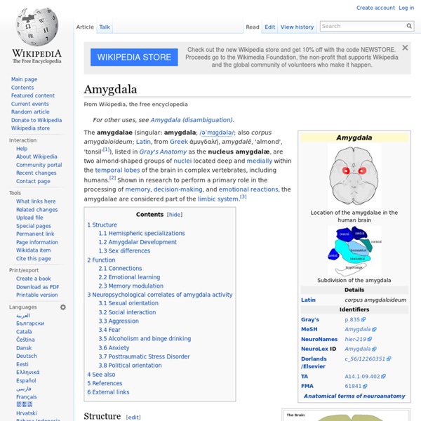
Limbic system The limbic system (or paleomammalian brain) is a complex set of brain structures that lies on both sides of the thalamus, right under the cerebrum.[1] It is not a separate system, but a collection of structures from the telencephalon, diencephalon, and mesencephalon.[2] It includes the olfactory bulbs, hippocampus, amygdala, anterior thalamic nuclei, fornix, columns of fornix, mammillary body, septum pellucidum, habenular commissure, cingulate gyrus, Parahippocampal gyrus, limbic cortex, and limbic midbrain areas. The limbic system supports a variety of functions, including emotion, behavior, motivation, long-term memory, and olfaction.[3] It appears to be primarily responsible for emotional life, and it has a great deal to do with the formation of memories. Structure[edit] The limbic system is the set of brain structures that forms the inner border of the cortex. The limbic system is made up of the limbic lobe and other deep-lying structures. Function[edit] Hippocampus[edit] Learning[edit]
Parahippocampal gyrus Parahippocampal gyrus It has been involved in some cases of hippocampal sclerosis.[2] Asymmetry has been observed in schizophrenia.[3] Structure[edit] The anterior part of the gyrus includes the perirhinal and entorhinal cortices[citation needed]. The term parahippocampal cortex is used to refer to an area that encompasses both the posterior parahippocampal gyrus and the medial portion of the fusiform gyrus. Function[edit] Scene recognition[edit] Damage to the PPA (for example, due to stroke) often leads to a syndrome in which patients cannot visually recognize scenes even though they can recognize the individual objects in the scenes (such as people, furniture, etc.). Social context[edit] Additional research has increased the probability that the right parahippocampal gyrus in particular has functions beyond the contextualizing of visual background. Additional images[edit] Animation. References[edit] External links[edit]
Parietal lobe The parietal lobe is one of the four major lobes of the cerebral cortex in the brain of mammals. The parietal lobe is positioned above (superior to) the occipital lobe and behind (posterior to) the frontal lobe and central sulcus. The parietal lobe integrates sensory information among various modalities, including spatial sense and navigation (See proprioception), the main sensory receptive area for the sense of touch (See somatosensation) in the somatosensory cortex which is just posterior to the central sulcus in the postcentral gyrus,[1] and the dorsal stream of the visual system. The major sensory inputs from the skin (touch, temperature, and pain receptors) relay through the thalamus to parietal lobe. Several portions of the parietal lobe are important in language processing. The name comes from the overlying parietal bone, which is named from the Latin paries-, "wall". Structure[edit] Animation. The parietal lobe is defined by three anafissure divides the two hemispheres.
Supplementary motor area Some motor areas in the human cortex. The supplementary motor area is shown in pink. Image by: Paskari The supplementary motor area (SMA) is a part of the primate cerebral cortex that contributes to the control of movement. It is located on the midline surface of the hemisphere just in front of (anterior to) the primary motor cortex leg representation. For the discovery of the SMA and its relationship to other motor cortical areas, see the main article on the motor cortex. Subregions[edit] At least six areas are now recognized within the larger region once defined as the SMA. The supplementary eye field (SEF) is a relatively anterior portion of the SMA that, when stimulated, evokes head and eye movements and perhaps movements of the limbs and torso.[8][9][10][11] SMA proper in monkeys has now been confined to a region on the crown of the hemisphere and extending partly onto the medial wall, just anterior to the primary motor leg representation. Functions[edit] References[edit]