
MicroRNA. Diagram of miRNA action with mRNA miRNAs are well conserved in eukaryotic organisms and are thought to be a vital and evolutionarily ancient component of genetic regulation.[9][10][11][12] While core components of the microRNA pathway are conserved between plants and animals, miRNA repertoires in the two kingdoms appear to have evolved independently with different modes of function.[13] Plant miRNAs usually have perfect or near-perfect pairing with their messenger RNA targets and induce gene repression through degradation of their target transcripts.[14][15] Plant miRNAs may bind their targets in both coding regions and untranslated regions.[15] In contrast, animal miRNAs are able to recognize their target mRNAs by as little as 6-8 nucleotides (the seed region) at the 5' end of an animal miRNA.[6][16] Combinatorial regulation is a feature of miRNA regulation.

History[edit] Nomenclature[edit] Biogenesis[edit] MicroRNAs are produced from either their own genes or from introns. X chromosome. Nucleus of a female amniotic fluid cell.

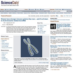
Top: Both X-chromosome territories are detected by FISH. Shown is a single optical section made with a confocal microscope. Bottom: Same nucleus stained with DAPI and recorded with a CCD camera. And it's all down to X-chromosome related microRNA. As anyone familiar with the phrase 'man-flu' will know women consider themselves to be the more robust side of the species when it comes to health and illness.
Now new research, published in BioEssays, seems to support the idea. The research focuses on the role of MicroRNAs encoded on the X chromosome to explain why women have stronger immune systems to men and are less likely to develop cancer. The research, led by Dr Claude Libert from Ghent University in Belgium, focused on MicroRNA, tiny strains of ribonucleic acid which alongside DNA and proteins, make up the three major macromolecules that are essential for all known forms of life. "Statistics show that in humans, as with other mammals, females live longer than males and are more able to fight off shock episodes from sepsis, infection or trauma," said Libert. Craniosynostosis. Craniosynostosis occurs in one in 2000 births.

New hope for children with craniosynostosis: Developing technologies to improve the treatment for premature fusion of skull bones in children. Cerebrospinal fluid. Vials containing human cerebrospinal fluid.

Cerebrospinal fluid (CSF) is a clear colorless bodily fluid found in the brain and spine. It is produced in the choroid plexus of the brain. It acts as a cushion or buffer for the brain's cortex, providing a basic mechanical and immunological protection to the brain inside the skull, and it serves a vital function in cerebral autoregulation of cerebral blood flow. Structure[edit] CSF circulation. Development of hydrocephalus and classical hypothesis of cerebrospinal fluid hydrodynamics. Spinal fusion. Spinal fusion, also known as spondylodesis or spondylosyndesis, is a surgical technique used to join two or more vertebrae.

Supplementary bone tissue, either from the patient (autograft) or a donor (allograft), is used in conjunction with the body's natural bone growth (osteoblastic) processes to fuse the vertebrae. Neurosurgeons use adult stem cells to grow neck vertebrae. Neurosurgery researchers at UC Davis Health System have used a new, leading-edge stem cell therapy to promote the growth of bone tissue following the removal of cervical discs -- the cushions between the bones in the neck -- to relieve chronic, debilitating pain.

The procedure was performed by associate professors of neurosurgery Kee Kim and Rudolph Schrot. It used bone marrow-derived adult stem cells to promote the growth of the bone tissue essential for spinal fusion following surgery, as part of a nationwide, multicenter clinical trial of the therapy. Removal of the cervical disc relieves pain by eliminating friction between the vertebrae and/or nerve compression. Spinal fusion is used following surgery for degenerative disc disease, where the cushioning cartilage has worn away, leaving bone to rub against bone and herniated discs, where the discs pinch or compress nerves. "For the past 50 years, bone marrow-derived stem cells have been used to rebuild patients' blood-forming systems. Adermatoglyphia. Adermatoglyphia is a rare medical condition which causes a person to have no fingerprints.

There are only four known extended families worldwide which are affected by this condition. Mutation linked with the absence of fingerprints. Scientists have identified a mutation that might underlie an extremely rare condition, called "adermatoglyphia," which causes people to be born without any fingerprints.

The research, published by Cell Press online August 4th in The American Journal of Human Genetics, not only provides valuable insight into the genetic basis of adermatoglyphia and of typical fingerprint formation but also underscores the usefulness of rare genetic mutations as a tool for investigating unknown aspects of our biology. Human skin has ridges called dermatoglyphs that are present on the fingers, palms, toes and soles. The dermatoglyphs on the finger tips, better known as fingerprints, are often used as a means for establishing identity.
Shake Hands with the Invisible Man. Shake Hands with the Invisible ManMonday, September 19, 2011 TAU researcher identifies genetic defect that leaves some without fingerprints Like DNA, fingerprints are unique to each person or set of identical twins.

That makes them a valuable identification tool for everything from crime detection to international travel.