
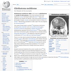
Glioblastoma multiforme. Glioblastoma multiforme (GBM), also known as glioblastoma and grade IV astrocytoma, is the most common and most aggressive cancer that begins within the brain.[1] Signs and symptoms are initially non-specific.
They may include headaches, personality changes, nausea, and symptoms similar to those of a stroke.[2] Worsening of symptoms is often rapid. This can progress to unconsciousness.[3] The cause of most cases is unclear.[3] Uncommon risk factors include genetic disorders such as neurofibromatosis and Li Fraumeni syndrome and previous radiation therapy.[3][4] Glioblastomas represent 15% of brain tumors.[2] They can either start from normal brain cells or develop from an already existing low-grade astrocytoma.[5] The diagnosis is typically made by a combination of CT scan, MRI scan, and tissue biopsy.[2] There is no clear way to prevent the disease. Signs and symptoms[edit] Risk factors[edit] Other risk factors include:[27] Pathogenesis[edit] Molecular alterations[edit]
Promising target found in treating deadly brain cancer. Researchers at the University of Virginia Cancer Center have identified a promising target for treating glioblastoma, one that appears to avoid many of the obstacles that typically frustrate efforts to develop effective treatments for this deadliest of cancers.

Glioblastoma multiforme. New 'scarless' surgery takes out tumors through natural skull opening. A technique developed by Johns Hopkins surgeons is providing a new route to get to and remove tumors buried at the base of the skull: through the natural hole behind the molars, above the jawbone and beneath the cheekbone.

In a report detailing the novel surgery, published in the October 2011 issue of The Laryngoscope, the surgeons say the procedure, already performed in seven patients, yields faster recovery and fewer complications than traditional approaches. And, because the incisions are made inside the cheek, there are no visible scars. Kofi Boahene, M.D., an assistant professor of facial plastic and reconstructive surgery and otolaryngology-head and neck surgery at the Johns Hopkins University School of Medicine, says the idea for the new approach came to him when a 20-year-old female patient previously treated for a brain tumor developed a new tumor deep in the skull base.
Not all patients are candidates for this procedure, Boahene cautions. Oligodendroglioma. Oligodendrogliomas are a type of glioma that are believed to originate from the oligodendrocytes of the brain or from a glial precursor cell.
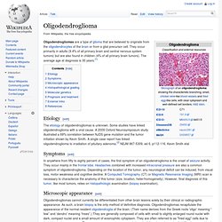
They occur primarily in adults (9.4% of all primary brain and central nervous system tumors) but are also found in children (4% of all primary brain tumors). The average age at diagnosis is 35 years.[1] How normal cells become brain cancers. Brain tumor specimens taken from neurosurgery cases at the University of California, San Francisco (UCSF) Medical Center have given scientists a new window on the transformation that occurs as healthy brain cells begin to form tumors.

The work may help identify new drugs to target oligodendroglioma, a common type of brain tumor, at its earliest stage, when it is generally most treatable. Any potential drugs identified will have to prove safe and effective in clinical trials, a process that can take several years. As described in the journal Cancer Cell this month, the UCSF team found that the pool of cells from which oligodendroglioma tumors emerge normally divide "asymmetrically" by splitting into two unequal parts -- like giving birth to fraternal twins who look different and have distinct fates. When these normal cells transform into cancer cells, they switch gears and begin dividing symmetrically, essentially giving birth to identical twins instead. Why Divisions Matter to Cancer.
Medulloblastoma. Medulloblastoma is a highly malignant primary brain tumor that originates in the cerebellum or posterior fossa.
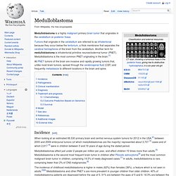
Tumors that originate in the cerebellum are referred to as infratentorial because they occur below the tentorium, a thick membrane that separates the cerebral hemispheres of the brain from the cerebellum. Another term for medulloblastoma is infratentorial primitive neuroectodermal tumor (PNET). New research method to identify brain tumors could enhance neurosurgery. The use of a new brain tumor-targeting contrast agent that differentiates between normal and cancer cells in conjunction with a high-powered microscopy system could potentially lead to a method of more precise neurosurgery for brain tumors, according to research paper published as a cover story in the December issue of Translational Oncology.

Developed by researchers in the Department of Biomedical Engineering (BME) at Stony Brook University, the contrast agent adheres to a molecular marker of medulloblastoma, a form of brain cancer, and can be seen by the optical microscope system, also developed by the research team. In their article entitled "Microscopic Delineation of Medulloblastoma Margins in a Transgenic Mouse Model Using a Topically Applied VEGFR-1 Probe," Stony Brook researchers Dr. Jonathan T.C. Liu, Assistant Professor of Biomedical Engineering, and BME graduate students Danni Wang, Steven Y. To further advance this hand-held microscopy and imaging technology, Dr. Could targeting a virus treat a common pediatric brain tumor? Public release date: 26-Sep-2011 [ Print | E-mail Share ] [ Close Window ] Contact: Karen Honeypress_releases@the-jci.org 734-546-5242Journal of Clinical Investigation Medulloblastomas are the most common cancerous (malignant) brain tumors in children.

Although survival rates have improved over the years, medulloblastoma remains associated with substantial mortality, and long-term survivors often suffer debilitating effects from the intensive treatments. A team of researchers, led by Cecilia Söderberg-Nauclér and John Inge Johnsen, at the Karolinska Institutet, Sweden, has now identified a potential target for a more cancer-specific approach to treating medulloblastoma that they hope could improve patient outcome.
Brain tumor. New tool to help surgeons remove more cancer tissue during brain surgery. Scientists are reporting development and successful initial testing of a new tool that tells whether brain tissue is normal or cancerous while an operation is underway, so that surgeons can remove more of the tumor without removing healthy tissue, improving patients' survival.

The report appears in ACS' journal Analytical Chemistry. Zoltán Takáts and colleagues point out that cancer can recur if tumor cells remain in the body after surgery. As a precaution, surgeons typically remove extra tissue surrounding a breast, prostate and other tumors in the body. But neurosurgeons face severe limitations because removing extra tissue can impair the patient's memory, mobility and other vital functions. Neurosurgeons thus strive to precisely identify the tumor margins during brain surgery.
Tagging tumors with gold: Scientists use gold nanorods to flag brain tumors. "It's not brain surgery" is a phrase often uttered to dismiss a job's difficulty, but when the task actually is removing a brain tumor, even the slightest mistake could have serious health consequences.
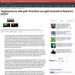
To help surgeons in such high-pressure situations, researchers from Prof. Adam Wax's team at Duke University's Fitzpatrick Institute for Photonics and Biomedical Engineering Department have proposed a way to harness the unique optical properties of gold nanoparticles to clearly distinguish a brain tumor from the healthy, and vital, tissue that surrounds it. The team will present their findings at the Optical Society's (OSA) Annual Meeting, Frontiers in Optics (FiO) 2011, taking place in San Jose, Calif. next week.
Current techniques for outlining brain tumors vary, but all have limitations, such as the inability to perform real-time imaging without big, expensive equipment, or the toxicity and limited lifespan of certain labeling agents.