
UW Histology homepage. Histology Website Resources Virtual Histology Loyola Univ Medical School.
LUMEN Histology home page. Histology Text Book Atlas & Histology Teaching Series on DVD Video. 26 DVD Lecture Course With Lab Material. Online Text-Atlas. 24 Power Point Programs. Complete Integrated Histology Course. SIU SOM Histology INTRO. How should I study histology?
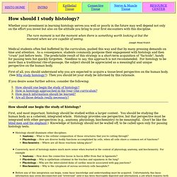
Whether your investment in learning histology serves you well or poorly in the future may well depend not only on the effort you invest but also on the attitude you bring to your first encounters with this discipline. Medical students often feel buffetted by the curriculum, pushed this way and that by many pressing demands on time and attention. As a consequence, students commonly postpone their engagement with histology and then "cram" just before tests. The predictable result of this strategy is a short-term acquisition of "factoids", handy for passing tests but quickly forgotten. Needless to say, this approach is not recommended.
A broad-based appreciation of Histology is most directly applicable to topics which are deemphasized in the Year One curriculum -- inflammation, tissue repair, and neoplasm. Sooner or later you will almost certainly wish you had learned more. But remember, don't try to learn more than you can understand! SIU SOM Histology. JayDoc HistoWeb. The Microscope-Blue Histology. Blue Histology - Nervous Tissue. Additional Resources These links will open a new browser window.
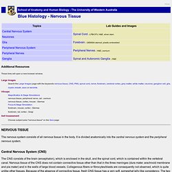
Large Images VScope Magnification & Stage Simulations nervous tissue, peripheral nerve, cat - osmium nervous tissue, cortex, mouse - Giemsa Focus & Stage Simulation: forebrain, mouse, cortex - Giemsa forebrain, rat, cortex - Golgi Self Assessment Choose subject area "nervous tissue" on the Quiz page The nervous system consists of all nervous tissue in the body. Central Nervous System (CNS) The CNS consists of the brain (encephalon), which is enclosed in the skull, and the spinal cord, which is contained within the vertebral canal.
Neurones The vast majority of neurones is generated before birth. Neurones are generally large cells. The shape and orientation of the dendritic tree of the neurone determines the amount and type of information that may reach the neurone. Transmitters Neurotransmitters either excite or inhibit the postsynaptic neurone. Receptors Suitable Slides Glia Many glial cells do express neurotransmitter receptors. Blue Histology - Muscle. Motion, as a reaction of multicellular organisms to changes in the internal and external environment, is mediated by muscle cells. The basis for motion mediated by muscle cells is the conversion of chemical energy (ATP) into mechanical energy by the contractile apparatus of muscle cells. The proteins actin and myosin are part of the contractile apparatus. The interaction of these two proteins mediates the contraction of muscle cells. Actin and myosin form myofilaments arranged parallel to the direction of cellular contraction.
A further specialisation of muscle cells is an excitable cell membrane which propagates the stimuli which initiate cellular contraction. Three structurally and functionally distinct types of muscle are found in vertebrates: smooth muscle , skeletal muscle and. Blue Histology - Connective Tissues. Additional Resources These links will open a new browser window.
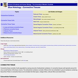
Large Images VScope Magnification & Stage Simulations dense irregular connective tissue, human, H&E - non-lactating breast reticular connective tissue, reticulin - liver Focus & Stage Simulations spleen, reticulin - reticular connective tissue Self Assessment Choose subject area "connective tissue" on the Quiz page. Connective tissue consists of cells separated by varying amounts of extracellular substance. Extracellular Substance Collagen fibres Collagen fibres are the dominant fibre type in most connective tissues. Epithelia and Glands- Blue Histology. Additional Resources These links will open a new browser window.
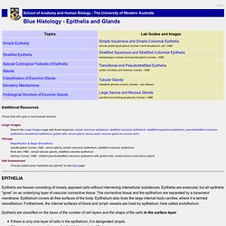
Large Images VScope Magnification & Stage Simulations: parotid gland, human, H&E - serous gland, simple columnar epithelium, stratified columnar epithelium thick skin, H&E - simple tubular glands, stratified cuboidal epithelium trachea, human, H&E - ciliated pseudostratified columnar epithelium with goblet cells, mixed mucous and serous gland Self Assessment Choose subject area "epithelia and glands" on the Quiz page Epithelia are tissues consisting of closely apposed cells without intervening intercellular substances. Epithelia are classified on the basis of the number of cell layers and the shape of the cells in the surface layer.
If there is only one layer of cells in the epithelium, it is designated simple. Simple Epithelia Simple squamous epithelium This type is composed of a single layer of flattened, scale- or plate-like cells. Simple cuboidal epithelium.