
TWIST1 Gene - GeneCards. Aliases for TWIST1 Gene Twist Family BHLH Transcription Factor 1 2 3 Twist Basic Helix-Loop-Helix Transcription Factor 1 2 3 Class A Basic Helix-Loop-Helix Protein 38 3 4 H-Twist 3 4 BHLHa38 3 4 TWIST 3 4 Blepharophimosis, Epicanthus Inversus And Ptosis 3 2 Twist Homolog 1 (Drosophila) 2 TWIST Homolog Of Drosophila 3 B-HLH DNA Binding Protein 3 Saethre-Chotzen Syndrome 2 Acrocephalosyndactyly 3 2 Craniosynostosis 2 Twist Homolog 1 3 BPES2 3 BPES3 3 CRS1 3 ACS3 3 CSO 3 SCS 3 CRS 3 External Ids for TWIST1 Gene Previous HGNC Symbols for TWIST1 Gene Previous GeneCards Identifiers for TWIST1 Gene Basic helix-loop-helix (bHLH) transcription factors have been implicated in cell lineage determination and differentiation.

Acts as a transcriptional regulator. No data available for Tocris Summary , PharmGKB "VIP" Summary , fRNAdb sequence ontologies and piRNA Summary for TWIST1 Gene. Mesenchymal Tissues. Muscle: There are three types of muscle: skeletal muscle, cardiac muscle, and smooth muscle.
The first two types are both forms of striated muscle.Skeletal muscle is called "voluntary" muscle because you make a conscious decision to move such muscles to perform some act, such as picking up a pencil or walking. A muscle is composed of individual muscle cells that contain many muscle filaments (myofibrils). The filaments compose a muscle fiber. A skeletal muscle fiber has multiple nuclei at its periphery. Vein. Veins are blood vessels that carry blood toward the heart.
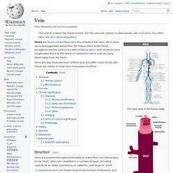
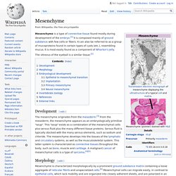
Most veins carry deoxygenated blood from the tissues back to the heart; exceptions are the pulmonary and umbilical veins, both of which carry oxygenated blood to the heart. In contrast to veins, arteries carry blood away from the heart. Structure[edit] Veins are present throughout the body as tubes that carry blood back to the heart. Veins are classified in a number of ways, including superficial vs. deep, pulmonary vs. systemic, and large vs. small. Mesenchymal tissue. Mesenchyme. Mesenchyme is a type of connective tissue found mostly during development of the embryo.[1] It is composed mainly of ground substance with few cells or fibers.
It can also be referred to as a group of mucoproteins found in certain types of cysts (etc.), resembling mucus. It is most easily found as a component of Wharton's jelly. CONNECTIVE TISSUE - BLOOD BLOODFORMING TISSUES. Blood is considered a connective tissue for two basic reasons: (1) embryologically, it has the same origin (mesodermal) as do the other connective tissue types and (2) blood connects the body systems together bringing the needed oxygen, nutrients, hormones and other signaling molecules, and removing the wastes.
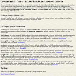
In circulating blood two different cell types are found: enucleated erythrocytes or red blood cells and nucleated leukocytes or white blood cells. We will study their histology in blood smears. Erythrocytes (red blood cells) RBCs are small (7 um) cells lacking a nucleus. They stain red with eosin and due to their concave shape have a lighter staining center. Leukocytes (white blood cells) The WBCs are divided into two groups: (1) granular leukocytes, containing distinctive cytoplasmic granules, including neutrophils, eosinophils and basophils and (2) agranular leukocytes, without granules, including monocytes and lymphocytes. Review. Epithelium. Epithelial layers contain no blood vessels, so they must receive nourishment via diffusion of substances from the underlying connective tissue, through the basement membrane.[1][2] Cell junctions are well-employed in epithelial tissues.
Classification[edit] Summary showing different epithelial cells/tissues and their characteristics. The three principal shapes associated with epithelial cells are—squamous, cuboidal and columnar. Blood - Embryonic Development & Stem Cells - LifeMap Discovery. Embryonic Development of the Blood: Vertebrate blood development occurs in two phases.
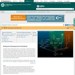
A transient embryonic (“primitive”) phase of hematopoiesis, beginning on E7.5 in the mouse embryo, is followed by the definitive (“adult”) phase, which begins on E9.5 in the liver of the developing mouse embryo. The embryonic phase of hematopoiesis is thought to initiate the circulation that provides the embryo with its initial blood cells and with its capillary network connecting it to the yolk. The definitive phase of hematopoiesis generates more cell types and provides the stem cells that will last throughout the vertebrate's lifetime. The Embryo Project Encyclopedia. Mesenchyme is a type of animal tissue comprised of loose cells embedded in a mesh of proteins and fluid, called the extracellular matrix.
The loose, fluid nature of mesenchyme allows its cells to migrate easily and play a crucial role in the origin and development of morphological structures during the embryonic and fetal stages of animal life. Mesenchyme directly gives rise to most of the body’s connective tissues, from bones and cartilage to the lymphatic and circulatory systems.
Furthermore, the interactions between mesenchyme and another tissue type, epithelium, help to form nearly every organ in the body. Although most mesenchyme derives from the middle embryological germ layer, the mesoderm, the outer germ layer known as the ectoderm also produces a small amount of mesenchyme from a specialized structure called the neural crest. Mesenchyme is generally a transitive tissue; while crucial to morphogenesis during development, little can be found in adult organisms. In 1888, N.