
Endometrial Cups and Secretion of Equine Chorionic Gonadotropin. Shortly after establishment of pregnancy in equids, high concentrations of the hormone equine chorionic gonadotropin (eCG) appear in the mare's serum.
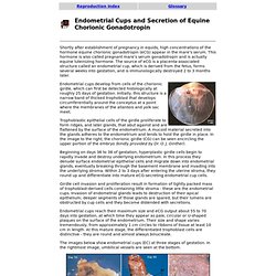
This hormone is also called pregnant mare's serum gonadotropin and is actually equine luteinizing hormone. The source of eCG is a placenta-associated structure called an endometrial cup, which is derived from the fetus, forms several weeks into gestation, and is immunologically destroyed 2 to 3 months later. Endometrial cups develop from cells of the chorionic girdle, which can first be detected histologically at roughly 25 days of gestation. Initially, this structure is a narrow band of thicked trophoblast that develops circumferentially around the conceptus at a point where the membranes of the allantois and yolk sac meet. Exercise 29 Avian Female Reproductive System. The Codfish lays ten thousand eggs, The Chicken lays but one; But a Codfish never cackles to tell you what she's done.
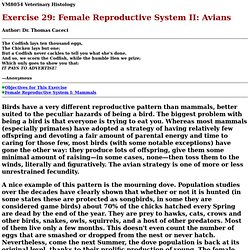
And so, we scorn the Codfish, while the humble Hen we prize; Which only goes to show you that:IT PAYS TO ADVERTISE! —Anonymous Objectives for This Exercise Female Reproductive System I: Mammals Birds have a very different reproductive pattern than mammals, better suited to the peculiar hazards of being a bird. A nice example of this pattern is the mourning dove. Avian Reproductive Pattern Avian reproduction has been best studied in the domestic chicken and turkeys, and the modern poultry industry rests on its complete understanding of the cycle and how to control it.
Birds lay eggs in clutches. In hens, ovulation usually occurs in the morning, and almost never after 3:00 PM under normal daylight conditions. The Reproductive Tract Ovary As with mammals, eggs are produced in ovaries, or, more correctly speaking, one ovary. To see the avian ovary click here. Infundibulum. Placental Structure and Classification.
The placentas of all eutherian (placental) mammals provide common structural and functional features, but there are striking differences among species in gross and microscopic structure of the placenta.
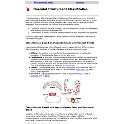
Two characteristics are particularly divergent and form bases for classification of placental types: The gross shape of the placenta and the distribution of contact sites between fetal membranes and endometrium.The number of layers of tissue between maternal and fetal vascular systems. Differences in these two properties allow classification of placentas into several fundamental types. Classification Based on Placental Shape and Contact Points Examination of placentae from different species reveals striking differences in their shape and the area of contact between fetal and maternal tissue: Diffuse: Almost the entire surface of the allantochorion is involved in formation of the placenta. Classification Based on Layers Between Fetal and Maternal Blood. Classification of Vaginal Epithelial Cells. A majority of cells observed in a normal vaginal smear are, not surprisingly, vaginal epithelial cells.
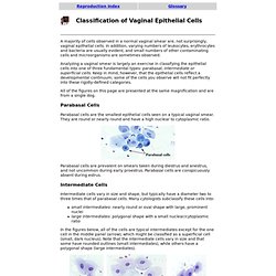
In addition, varying numbers of leukocytes, erythrocytes and bacteria are usually evident, and small numbers of other contaminating cells and microorganisms are sometimes observed. Analyzing a vaginal smear is largely an exercise in classifying the epithelial cells into one of three fundamental types: parabasal, intermediate or superficial cells. Keep in mind, however, that the epithelial cells reflect a developmental continuum; some of the cells you observe will not fit perfectly into these rigidly-defined categories. All of the figures on this page are presented at the same magnification and are from a single dog. Parabasal Cells Parabasal cells are the smallest epithelial cells seen on a typical vaginal smear.
Cytologic Changes Through the Canine Estrous Cycle. Stages of the canine estrous cycle can be defined by sexual behavior, physical signs (vulvar swelling, vaginal bleeding) or by vaginal cytology.
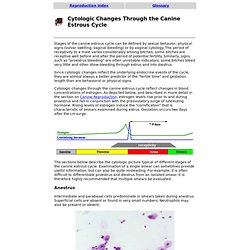
The period of receptivity to a male varies considerably among bitches; some bitches are receptive well before and after the period of potential fertility. Similarly, signs such as "proestrus bleeding" are often unreliable indicators; some bitches bleed very little and other show bleeding through estrus and into diestrus. Since cytologic changes reflect the underlying endocrine events of the cycle, they are almost always a better predictor of the "fertile time" and gestation length than are behavioral or physical signs.
Cytologic changes through the canine estrous cycle reflect changes in blood concentrations of estrogen. As depicted below, and described in more detail in the section on Canine Reproduction, estrogen levels rise prior to and during proestrus and fall in conjunction with the preovulatory surge of luteinizing hormone. Anestrus Estrus.