
The Breast. Veins The blood from the upper limb is returned to the heart by two sets of veins, superficial and deep.
Both sets have valves, and both drain ultimately into the axillary vein. Superficial Veins (fig. 7-1). Histology images of Breast by PathPedia.com: Pathology e-Atlas. [NORMAL BREAST HISTOLOGY].
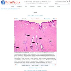
Breast can be considered a modified skin appendage in Mammalians producing milk for the nourishment of the newborn. The milk is discharged from a collection of 10-20 large ducts opening through pores in the nipple during lactation. Each large duct, also called lactiferous duct, branches out deep into the breast tissue forming what is called a breast lobe. Thus, a breast consists of about 10-20 inter-anastomosing lobes separated from each other by varying amounts of fibro-adipose tissue. This photomicrograph shows six (6) lactiferous ducts (arrows) that run from the nipple to branch down into successive smaller ducts until the formation of “terminal duct lobular unit (TDLU).” [NORMAL BREAST HISTOLOGY]. [NORMAL BREAST HISTOLOGY]. [NORMAL BREAST HISTOLOGY]. Embryology. Histology Guide. White blood cells White blood cells are much less common than red blood cells.
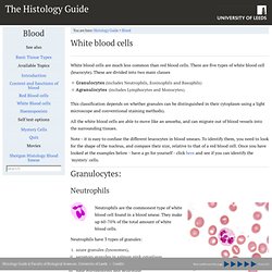
There are five types of white blood cell (leucocyte). These are divided into two main classes Granulocytes (includes Neutrophils, Eosinophils and Basophils) Agranulocytes (includes Lymphocytes and Monocytes). This classification depends on whether granules can be distinguished in their cytoplasm using a light microscope and conventional staining methods). All the white blood cells are able to move like an amoeba, and can migrate out of blood vessels into the surrounding tissues. Note - it is easy to confuse the different leucocytes in blood smears. Granulocytes: Neutrophils This shows a neutrophil in a blood smear. SKIN. Fig_3_2.gif (393×350) Cartilage. Dental Histology Fall 2003. Skeletal System : Bone Formation ( Intramembranous Ossification & Endochondral Ossification) CARTILAGE AND BONE CELLS. Groups of 1-4 chondrocytes in hyaline cartilage.
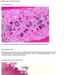
What are isogenous groups? Study the perichondrium, outside cartilage, find layers of flattened cells that may give rise to chondrocytes as they begin to produce matrix. These are called chondroblasts. They are responsible for appositional growth. Exercise 7: Cartilage. VM8054 Veterinary Histology Exercise 7 Author: Dr.
Thomas Caceci Objectives for This Exercise Connective tissues are those with supportive functions. Note that word "replaced. " Cartilage Tissue - Structure and Functions of Human Tissue Types. Note: This page is part of the section about the structure and function of different Tissue Types, which is related to the section about Histology and Cells (incl. structure of animal cells, cell division, mitosis, meiosis).
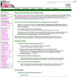
This "Tissue Types" section is included to complete description of the knowledge of "Histology - The Cell" required by some courses in First-Level Anatomy and Physiology. To read about other tissue types see the list of on the left. In the fetus and infant cartilage occurs in many parts of the body but much of this juvenile cartilage disappears during growth and development. The information on this page is concerned primarily with cartilage tissue in adult humans. Cartilage is a connective tissue consisting of a dense matrix of collagen fibres and elastic fibres embedded in a rubbery ground substance. What Is the Function of Compact Bone? Untitled Document. Practice Exam Connective Tissue. Answer the multiple choice question below each photo.
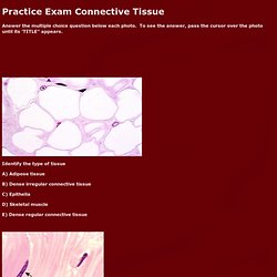
To see the answer, pass the cursor over the photo until its 'TITLE" appears. Identify the type of tissue A) Adipose tissue B) Dense irregular connective tissue C) Epithelia D) Skeletal muscle E) Dense regular connective tissue 2. A) Fat B) Histamine C) Antibodies D) Collagen fibers 3. A) Lymphocyte B) Mast cell C) Macrophage D) Plasma Cell. Histolab4e.htm. Cartilage and Bone, Demonstrations Go Back Hyaline Cartilage - Note glassy nature of the matrix.
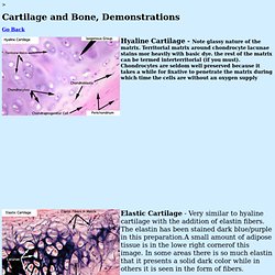
Territorial matrix around chondrocyte lacunae stains mor heavily with basic dye. the rest of the matrix can be termed interterritorial (if you must). Chondrocytes are seldom well preserved because it takes a while for fixative to penetrate the matrix during which time the cells are without an oxygen supply Elastic Cartilage - Very similar to hyaline cartilage with the addition of elastin fibers. Fibrocartilage - which is a combination of hyaline cart. and collagen type I. Microanatomy Web Atlas. The Gastrointestinal Immune System. The lumen of the gastrointestinal tract is outside of the body and much of it is heavily populated with potentially pathogenic microorganisms.
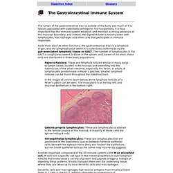
It is thus important that the immune system establish and maintain a strong presence at this mucosal boundary, and indeed, the digestive tube is heavily laden with lymphocytes, macrophages and other cells that participate in immune responses. Aside from all of its other functions, the gastrointestinal tract is a lymphoid organ, and the lymphoid tissue within it is collectively referred to as the gut-associated lymphoid tissue or GALT. The number of lymphocytes in the GALT is roughly equivalent to those in the spleen, and, based on location, these cells are distributed in three basic populations: Ligament and Tendon Structure and Function. BME 456: Biosolid Mechanics: Modeling and Applications General Info, Grading, Syllabus Instructor: Scott J.
Hollister, PhD Professor of Biomedical Engineering, Surgery, and Mechanical Engineering Announcements; Contact: Rm. 2208 LBME.