Brain Anatomy. Neurons. BrainImages. Spine. Drugs, Receptors. PeriNeuronalNet. Enteric NS. How Many Brain Cells? Radiology Key. Human Brain Anatomy in 3D. Lesion studies in contemporary neuroscience. Kainic acid. Human Brain Lestion Studies. Human Lesion Studies in the 21st Century. White Matter & Brain Volume. Introduction The human brain comprises both gray matter and white matter (WM), with the latter constituting roughly 60% of the total volume.
Gray matter consists of neuronal cell bodies, their dendrites and axons, glial cells, and blood vessels (1). On the other hand, WM consists of myelinated and unmyelinated axons that connect various gray matter areas of the brain and support communication between neurons, as well as convey information among the network of efferent and afferent axonal fibers. Disruption of these conduction pathways may cause motor and sensory dysfunction, neurobehavioral syndromes, and cognitive impairment (2–4). Well preserved 2,000-year-old brain cells found in Vesuvius victim. Brain cells have been found in exceptionally preserved form in the remains of a young man killed in the eruption of Mount Vesuvius almost 2,000 years ago, an Italian study has revealed.

The preserved neuronal structures in vitrified or frozen form were discovered at the archaeological site of Herculaneum, an ancient Roman city engulfed under a hail of volcanic ash after nearby Mount Vesuvius erupted in 79AD. “The study of vitrified tissue as the one we found at Herculaneum ... may save lives in future,” study lead author Pier Paolo Petrone, forensic anthropologist at Naples’ University Federico II, told the AFP news agency.
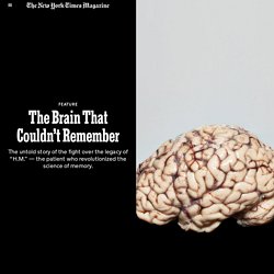
The Battle Over One Of The Most Studied Brains In Science. Aired 8/12/16 on KPBS News Researchers interested in how the brain creates new memories studied Henry Molaison, or "patient H.M.," who became perhaps the most famous research subject in recent history.

After Molaison died in 2008, his brain was moved to UC San Diego for further research. But it wouldn't stay there long. He was perhaps the most studied research subject in history, but he constantly forgot he was being studied. Known only as "patient H.M.” during his lifetime, Henry Molaison was unable to retain new memories for more than about 30 seconds. In 1953, at the age of 27, Molaison turned to a Connecticut neurosurgeon named William Scoville for help with his severe epilepsy. After his death, H.M.’s brain was moved to UC San Diego for further research. Dittrich wrote the book partly because he wanted to explore a dark chapter in his own family’s history. The Brain That Couldn’t Remember. In that pantheon of illuminatingly broken men and women, Henry stands apart.
It is difficult to exaggerate the impact he has had on our understanding of ourselves. Before Brenda Milner collaborated on that first paper about Henry, the prevailing theory of memory held that its functions could not be localized to a single cortical area, that learning was distributed across the brain as a whole. Brain and Society — THE BRAIN OBSERVATORY® Sebastian Seung: I am my connectome. A Human Memory Circuit Derived From Brain Lesions Causing Amnesia - PubMed.
A Connectomic Atlas of the Human Cerebrum—Chapter 6: The Temporal Lobe. Neuro3(2) Whole-Brain Vasculature Reconstruction at the Single Capillary Level. Animal models Four months old male mice from the C57 line were used for blood vessels tomographies and for two-photon comparison between Gel-BSA-FITC and lectin-FITC stained blood vessels.
For neuronal and blood vessels imaging of the same brain, a two-months old transgenic (Thy1-GFP-M line) male mouse expressing the Green Fluorescent Protein (GFP) in sparse pyramidal neurons was used38. All experimental protocols involving animals were designed in accordance with the laws of the Italian Ministry of Health. All experimental protocols were approved by the Italian Ministry of Health.
Blood-vessel lumen staining We used the protocol described in Tsai et al.21. Gel compositions Gel solutions were made of porcine skin gelatine type A (no. Vessel staining with lectin-FITC For SNR and segmentation performance comparison between gel-BSA-FITC and lectin-FITC, the lectin-FITC staining was performed via intracardial perfusion. Tissue transformation with CLARITY TDE clearing Image segmentation. Widespread Increased Diffusivity Reveals Early Cortical Degeneration in Huntington Disease.
Protein aggregation in cell biology: An aggregomics perspective of health and disease. The Structural and functional connectivity of the amygdala. Neural Encoding Spikes. FopOcxx.jpg (JPEG Image, 569 × 720 pixels) Scientists Surprised to Find No Two Neurons Are Genetically Alike. The past few decades have seen intensive efforts to find the genetic roots of neurological disorders, from schizophrenia to autism.

But the genes singled out so far have provided only sketchy clues. Even the most important genetic risk factors identified for autism, for example, may only account for a few percent of all cases. Much frustration stems from the realization that the key mutations elevating disease risk tend to be rare, because they are less likely to be passed on to offspring.
More common mutations confer only small risks (although those risks become more significant when calculated across an entire population). There are several other places to look for the missing burden of risk, and one surprising possible source has recently emerged—an idea that overturns a fundamental tenet of biology and has many researchers excited about a completely new avenue of inquiry. One question to be explored is whether genes associated with a brain disorder may harbor somatic mutations. UBC Undergraduate Medicine: Neuroanatomy. Bipartite Brain. And an associated story/painting The lower and upper modules differ in information processing "style".

Among other differences, the lower is capable of dealing productively with large numbers of variables with poorly specified relations among them and is capable of doing large numbers of tasks simultaneously while the upper the upper works better in terms of particular tasks which it tends to try and deal with in terms of smaller numbers of variables with well-defined and preferably uni-directional relations among them. The two modules have differing strengths and weaknesses. The upper module is distinctive in having the ability to notice and extend patterns and, using these, to conceive organizations other than those that can be derived from patterns of activity available to the lower module.
Myelination in Action! VBM.ashburner. Study sheds new light on brain's source of power. Department Of Psychiatry - Harvard Medical School - Research. The STUDY OF ADULT DEVELOPMENT The Study of Adult Development1 Division of Psychiatry, Brigham and Women's Hospital George E.
Vaillant, M.D..,;Vaillant, George E.,, Xing-jia Cui, M.D. 1432.full.pdf. About Us - Visionlearning.