
Ecole doctorale Biochimie et Biologie Moléculaire B2M. Dendritic cell. Dendritic cells (DCs) are antigen-presenting cells, (also known as accessory cells) of the mammalian immune system.
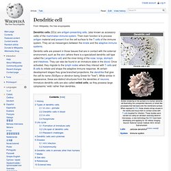
Their main function is to process antigen material and present it on the cell surface to the T cells of the immune system. They act as messengers between the innate and the adaptive immune systems. History[edit] Dendritic cells were first described by Paul Langerhans (hence "Langerhans cells") in the late nineteenth century. The term "dendritic cells" was coined in 1973 by Ralph M. Types of dendritic cells[edit] As in all dendritic cells, their particular morphology results in a very large contact surface to their surroundings compared to overall cell volume. Lymph node. Lymph nodes also have clinical significance.
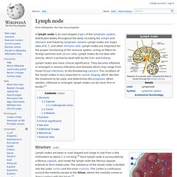
They become inflamed or enlarged in various infections and diseases which may range from trivial throat infections, to life-threatening cancers. The condition of the lymph nodes is very important in cancer staging, which decides the treatment to be used, and determines the prognosis. When swollen, inflamed or enlarged, lymph nodes can be hard, firm or tender.[1] Structure[edit] 1) Capsule; 2) Subcapsular sinus; 3) Germinal centre; 4) Lymphoid nodule; 5) Trabeculae Lymph nodes are bean or oval shaped and range in size from a few millimeters to about 1–2 cm long.[2] Each lymph node is surrounded by a fibrous capsule, and inside the lymph node the fibrous capsule extends to form trabeculae.
Thin reticular fibers and elastin form a supporting meshwork called a reticular network (RN) inside the node. The number and composition of follicles can change especially when challenged by an antigen, when they develop a germinal center.[2] Regulation of Dendritic Cell Migration by CD74, the MHC Class II-Associated Invariant Chain — Supporting Online Material. Déplacement cellule WT ((film) Myosine. Un article de Wikipédia, l'encyclopédie libre.
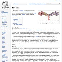
Portion de la structure de la myosine, les chaînes lourdes sont coloriées en rouge à gauche de l'image et les chaînes légères en jaune et orange Constitution[modifier | modifier le code] Elle est composée de deux chaînes lourdes (dimère) de 2 000 acides aminés dont les extrémités C-terminales en hélice α sont surenroulées, ce qui confère à la myosine la forme rigide d'un bâtonnet de 180 nm, et dont les têtes N-terminales de 190 acides aminés constituent les domaines moteurs. Sont associées à ces deux chaînes lourdes deux paires de chaînes légères : une paire de chaînes légères dites essentielles (ELC), et une paire de chaînes légères dites régulatrices (RLC) qui stabilisent la longue hélice près du domaine N-terminal (tête), dans la région du cou qui forme le ‘bras de levier’. Formes[modifier | modifier le code] L’expression de ces isoformes de chaîne lourde de myosine, dépend du type de fibre (rapide, lente) mais aussi de l’espèce.
Actin. A large number of illnesses and diseases are caused by mutations in alleles of the genes that regulate the production of actin or of its associated proteins.
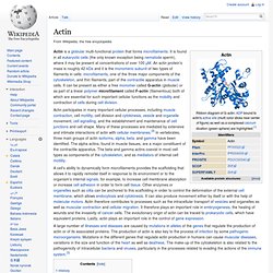
The production of actin is also key to the process of infection by some pathogenic microorganisms. Mutations in the different genes that regulate actin production in humans can cause muscular diseases, variations in the size and function of the heart as well as deafness. The make-up of the cytoskeleton is also related to the pathogenicity of intracellular bacteria and viruses, particularly in the processes related to evading the actions of the immune system.[3]
CD74. HLA class II histocompatibility antigen gamma chain also known as HLA-DR antigens-associated invariant chain or CD74 (Cluster of Differentiation 74), is a protein that in humans is encoded by the CD74 gene.[1][2] The invariant chain (Abbreviated Ii) is a polypeptide involved in the formation and transport of MHC class II protein.[3] The cell surface form of the invariant chain is known as CD74.
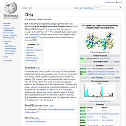
Function[edit] Possible interactions[edit] In limited cases, CD74 might interact with Macrophage migration inhibitory factor.[4] Déplacement cellule KO (film) LPTMC - Université Paris 6 - UPMC - CNRS. Microfluidics. Microfluidics is a multidisciplinary field intersecting engineering, physics, chemistry, biochemistry, nanotechnology, and biotechnology, with practical applications to the design of systems in which small volumes of fluids will be handled.
Microfluidics emerged in the beginning of the 1980s and is used in the development of inkjet printheads, DNA chips, lab-on-a-chip technology, micro-propulsion, and micro-thermal technologies. It deals with the behavior, precise control and manipulation of fluids that are geometrically constrained to a small, typically sub-millimeter, scale. Typically, micro means one of the following features: small volumes (µL, nL, pL, fL)small sizelow energy consumptioneffects of the micro domain Typically fluids are moved, mixed, separated or otherwise processed. Dendritic cell. 102, 058103 (2009): Pushing off the Walls: A Mechanism of Cell Motility in Confinement. We propose a novel mechanism of cell motility, which relies on the coupling of actin polymerization at the cell membrane to geometric confinement.
We consider a polymerizing viscoelastic cytoskeletal gel confined in a narrow channel, and show analytically that spontaneous motion occurs. Interestingly, this does not require specific adhesion with the channel walls, and yields velocities potentially larger than the polymerization velocity. The contractile activity of myosin motors is not necessary to trigger motility in this mechanism, but is shown quantitatively to increase the velocity. Our model qualitatively accounts for recent experiments which show that cells without specific adhesion proteins are motile only in confined environments while they are unable to move on a flat surface, and could help in understanding the mechanisms of cell migration in more complex confined geometries such as living tissues.
Mathematical Modelling of Natural Phenomena - Mechanisms of Cell Motion in Confined Geometries.