
Understanding Disease at the Organelle Level: created with Zunal WebQuest Maker. Task 2: Investigate the connections between dysfunctional organelles and disease.
A) Directions: The websites listed below contain information on various diseases and the dysfunctional organelles that cause them. Investigate these diseases. Fill-in the chart on your worksheet with the following information: (1) name of disease (2) organelle responsible for the disease (3) explain how the disease is a result of the malfunctioning organelle. B) Directions: Watch the YouTube video below and answer the prompt on the WebQuest worksheet. Prompt: One of the symptoms of mitochondrial disease is fatigue (weak and tired).
Task 3: Apply your knowledge of organelle structure and function to solve medical case studies. Directions: Choose 3 of the 4 case studies below. Case Study #1: George is an 18 year old male, who arrived in the emergency room (ER) at 6:03 a.m. with severe indigestion. Case Study #2: Lauren is a senior at a nearby high school. Case Study #3: Case Study #4: Hereditary elliptocytosis. Hereditary elliptocytosis, also known as ovalocytosis, is an inherited blood disorder in which an abnormally large number of the patient's erythrocytes (i.e. red blood cells) are elliptical rather than the typical biconcave disc shape.
It is one of many red-cell membrane defects. In its severe forms, this disorder predisposes to haemolytic anaemia. Although pathological in humans, elliptocytosis is normal in camelids. Historical perspective[edit] Elliptocytosis was first described in 1904,[1] and was first recognised as a hereditary condition in 1932.[2] More recently it has become clear that the severity of the condition is highly variable,[3] and there is much genetic variability amongst those affected.[4] Genetic prevalence[edit] There are three major forms of hereditary elliptocytosis: common hereditary elliptocytosis, spherocytic elliptocytosis and southeast Asian ovalocytosis.
The following categorisation of the disorder demonstrates its heterogeneity:[10] Pathophysiology[edit] Mitochondrial Disease Action Committee - MitoAction. What are Mitochondria?

Mitochondria are tiny organelles found in almost every cell in the body.They are known as the "powerhouse of the cell. "They are responsible for creating more than 90 percent of cellular energy.They are necessary in the body to sustain life and support growth.They are composed of tiny packages of enzymes that turn nutrients into cellular energy.Mitochondrial failure causes cell injury that leads to cell death. When multiple organ cells die there is organ failure.
Watch the 5-minute video, "How energy is made" back to top. What is Fabry Disease? By Sally Robertson, BSc Fabry disease is a rare genetic condition that belongs to a group of disorders referred to as lysosomal storage disease.

This condition is also called Anderson-Fabry disease, Fabry’s disease, angiokeratoma corporis diffusum and alpha-galactosidase A deficiency. The disease is named after a Johannes Fabry who discovered it. Disease inheritance Fabry disease is inherited in an X-linked manner, meaning the mutation that causes this condition is carried on the X chromosome. Fabry disease affects around 1 in 40,000 to 60,000 boys. Pathology and symptoms Fabry disease is caused by a deficiency in the enzyme alpha-galactosidase A, which used to be known of as ceramide trihexosidase. As the globotriaosylceramide accumulates, the blood vessels become increasingly narrow, preventing the flow of blood and nutrients to tissues that are usually supplied by the vessels.
Symptoms Symptoms of Fabry disease usually develop during childhood or adolescence. Treatment Prognosis. Animation - Endosymbiosis. The Virtual Cell. Table of Contents: Cell Biology. Cellcraft.
Build-a-Cell. Cell Size and Scale. Some cells are visible to the unaided eye The smallest objects that the unaided human eye can see are about 0.1 mm long.
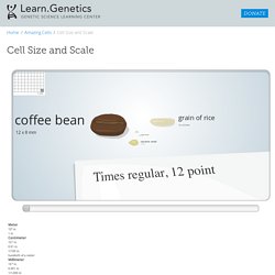
That means that under the right conditions, you might be able to see an ameoba proteus, a human egg, and a paramecium without using magnification. A magnifying glass can help you to see them more clearly, but they will still look tiny. Smaller cells are easily visible under a light microscope. It's even possible to make out structures within the cell, such as the nucleus, mitochondria and chloroplasts. To see anything smaller than 500 nm, you will need an electron microscope. Adenine The label on the nucleotide is not quite accurate. How can an X chromosome be nearly as big as the head of the sperm cell? No, this isn't a mistake. The X chromosome is shown here in a condensed state, as it would appear in a cell that's going through mitosis.
A chromosome is made up of genetic material (one long piece of DNA) wrapped around structural support proteins (histones). Carbon. Construction of the Cell Membrane. Inside a Cell.