
Imperative Considerations After Diagnosed With Basal Cell Carcinoma. Basal or squamous cell carcinoma is a type of skin cancer.
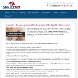
It begins in the basal cells- a type of cell within the skin that produces new skin as old ones die off. With early detection and treatment, almost all basal cell carcinoma(s) can be successfully removed without any complications. You just have to be proactive and if you see any change or unusual skin growths, consult with your dermatologist. Mobile Mohs Tech. Skin Cancer Types And Its Symptoms. Histopathology Services in California, USA. A specimen brought to the histopathology laboratory must first be logged, identified, and then subjected to specimen preparation prior to tissue processing.
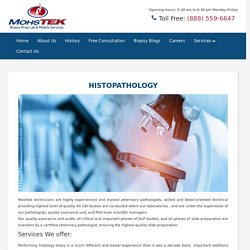
Sample collection Logging of specimen Preparation of tissues which include fixation and decalcification Processing of tissues includes dehydration, clearing, paraffin impregnation. Tissue embedding Preparation of sections which include the process of microtomy, attachment of sections to slide, and removal of pigments and precipitates Staining and mounting procedure, which includes dewaxing, hydration, staining dehydration, clearing, and mounting. Our state-of-the-art extensive experience and technology with biologics, antibodies, large molecules, small molecules, and devices help us to offer comprehensive pathologic assessments and interpretations. These are specialty pathology services provide you with the best flexible, cost-effective, and timely product-specific resources possible.
Guide to Dermatologic Histology Specimens Examination for Skin Treatment. No one wants to deal with acne, eczema, skin damage, scars, and other skin related diseases.
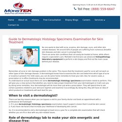
Yet around 50% of people are suffering from numerous dreadful skin diseases and skin cancer is amongst them. There are some skin conditions that can easily be treated at home, while others require a specialist’s care. Mohstek’s technicians use advanced histopathology laboratory equipment to perform a skin biopsy and find out the main cause behind your disease.
Remember all acne or skin damage problem is the same. This means that the treatment used for acne will not work on other types of skin damage disease. Histopathology Services In California. Histopathology Laboratory No Obligation for a Free Consultation!!!
For all of your Pathology needs, our Histology / Mohs laboratory and our mobile Mohs Technicians are ready to serve you. Fast, convenient, and flexible billing options are available, and always tailor fit the flow of every medical office. For over 25 years, our staff has been serving the medical community with the highest quality slides. Our Biopsy Prep Lab handles all 50 states located within the U.S. Forensic Pathology Services. Mohs Laboratory- Mohstek. Mobile Mohs Service Provider in the California, USA. Mohs Histology Laboratory. Routine Histology Lab managers, section leaders & supervisors monitor the histology aspects, ensuring that all the work is done according to explicit study conventions. All the activities experience basic stage examinations by our Quality Assurance Unit, which works autonomously of all venture exercises, guaranteeing consistency with GLP necessities.
Paraffin embedding, tissue processing, staining & cutting done with the help of histo-technicians & fully automated equipment with 10 years of experience. Amazing outcomes are ensured by our restrictive multi-step QC procedure to take out slides with tearing, wrinkles, blade artifacts, folds, non-uniform recoloring, foreign bodies & air bubbles. Glass slides are subjected to the latest digitized high-throughput scanner- Leica AT2. Immunohistochemistry using an automated machine. New antibody streamlining Exclusive conventions for immune system conditions not accessible at producer’s site, for example, PDL1 & CD8 for mouse tissue Safe. Mohs Laboratory- Mohstek. Histopathology Laboratory Equipment. Histopathology Laboratory Equipment. Histopathology Laboratory Equipment. Mohs technician- histology laboratory- Forensic pathology services.
Biopsy prep lab (2) Mobile Mohs technicians- Mobile lab services. Innovative Technologies In Dermatology. The Dermatology Industry is encountering maybe its most quick pace of development & innovation ever.
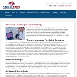
Naturally, this move has been driven to some extent by technology, with new advancements exhibiting tantalising value for healthy skin. Technology has transformed dermatology praxis which will accelerate in the years to come. Some of the digital technologies which help medical professionals truly bring dermatology into the 21st century are enlisted here- Tele-dermatology For Quick Response It is a win-win situation for everybody: there is no need to wait in the crowded rooms for an examination and dermatologists can manage the simple cases in shorter time online.Nobody appreciates spending hours in the clinic hanging tight for results. Innovative Technologies In Dermatology. Medical Laboratory Services. Biopsy blogs- Mohstek. Medical Laboratory Services. Mohs technician- histology laboratory- Forensic pathology services. Why Routine Histology is important. Histology, also called microanatomy or microscopic anatomy is the study of tissues and cells.
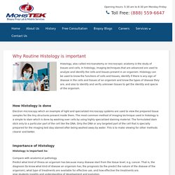
In histology, imaging techniques that are advanced are used to analyze and identify the cells and tissues present in an organism. Histology can be used to know the functions of cells and tissues, identify if there is any sign of disease in the cells and tissues of an organism and know the types of disease they are, and also to identify and verify unknown tissues to get the identity and specie of the organism. How Histology is done Electron microscopy which an example of light and specialized microscopy systems are used to view the prepared tissue samples for the tiny structures present inside them. The most common method of imaging technique used in histology is a simple to stain which is done by washing over cells by using highly specialized staining material. Histology for Beginners. The microscopic study of tissues and cells is known as histology.
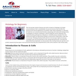
It includes concentrating on their unmistakable minuscule structure that clarifies the practical and clinical significance of sample under study. Histology is a vital part of medicine as it connects biochemistry, physiology and molecular biology to the investigation of ailment.Histology is used to diagnose various diseases and comprehending pathogenesis. Biopsies or unhealthy tissue samples from the influenced organs are generally handled, recoloured for observation under the microscope. Histology employs both electron microscopy & light microscopy, contingent upon the prerequisites of the sample being pictured.This field has extraordinarily progressed, as new practices like cloning,molecular genetics and protein sequencing have given more prominent learning and comprehension of the cells & tissues in the human body. Introduction to Tissues & Cells.
What is Mohs micrographic surgery and why it is considered more rather than other treatments? Are you suffering from skin lesion?
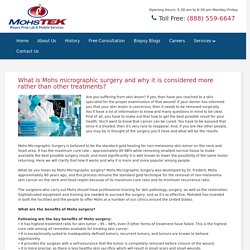
If yes, then have you reached to a skin specialist for the proper examination of that wound? If your doctor has informed you that your skin lesion is cancerous, then it needs to be removed surgically. You’ll have a lot of information to know and many questions in mind to be clear. First of all, you have to make out that how to get the best possible result for your health. You’ll want to know that cancer can be cured. Mohs Micrographic Surgery is believed to be the standard gold healing for non-melanoma skin tumor on the neck and head area. Mohs technician- histology laboratory- Forensic pathology services. Things You Should Know About Histopathology Laboratory. One of the most growing spectrums of science is histopathology.
It entails the evaluation of sampled whole tissues under the microscope. There are plenty of research organizations and companies that are dedicated to exploring this field and histopathology laboratory is one of them. Specimens that are required by the histopathology laboratory require preparation of tissues and examined using various techniques that are suitable for the kind of investigation and tissue required. Why Routine Histology is important. Benefits of Digital Pathology. Digital Pathology extends the limits of microscopy, allowing educators, students, clinicians, and students to share the tissue samples easily.
Image sent or shared through specific analysis software or over the internet paves the way to a new and exciting microscopy tool. Benefits of Digital Pathology People who utilize a microscope potentially leverage the digitized pathology images. Before the advent of this innovation, histological slides and photographs were the primary ways to share the images seen under a microscope with others. However, Digital Pathology eliminates issues related to sharing slides like degradation of samples and the inability to share samples of live cells.
Diagnostics – A hospital can send images globally, probably reducing the actual time it usually takes to diagnose and treat a pathogen. Education – Colleges or universities can access a massive database of samples online or via a network database, thus saving money for histological slides. Histology Laboratory. Histopathology and Histology are often combined to understand the histopathological interpretation better. It is essential to prepare the histology slides of a sample in the histology laboratory and evaluate them in the first place to determine if the cells or tissue are in a good state or diseased. Past In the 19th century, histology was a well-known academic discipline, and the first half of the 20th century was a productive period for staining procedures in histology and histopathology. Various centenary staining techniques are still being used and continue to offer valuable diagnostic details.
The first microscope was constructed in 1591; however, it had many optical issues. Things You Should Know About Histopathology Laboratory. Biopsy blogs- Mohstek. Mobile Mohs technicians- Mobile lab services. Use our mobile lab services for immediate answers on-site. Our mobile Mohs services are the fastest and the most accurate. The mobile lab services help you to prepare frozen section slides.
Our mobile lab services are delivered by local traveling technicians in your area. A full team of travel MOHS technicians are ready to serve you. They come prepared with all of the supplies needed. We have: Dermatologic Histology Specimens. Major Benefits of Mohs Micrographic Surgery Procedure for Skin Cancer. Undoubtedly, Mohs micrographic surgery procedure has the highest cure rate among skin cancer treatment modalities.
Recent studies have shown that Mohs surgery can have a 99% cure rate for many skin cancers. Benefits of Digital Pathology and Its Future Outlook. Major Benefits of Mohs Micrographic Surgery Procedure for Skin Cancer. Skin cancer surgery on face. Mobile Mohs technicians — A Detailed Guide on Mohs Micrographic Surgery. The Rising Trend of Digital Pathology in the World of Technology. The Histopathology laboratory equipment procedure involves examination of sampled tissues under a microscope using several kinds of specimens. They are larger specimens that include organs which are removed during a surgical operation. Pieces of tissue are a test which involves pieces of tissues that are taken to perform the test.
Fluid and tiny pieces of tissues can be obtained through a fine needle aspiration (FNA). Uses of Histopathology • The samples of the tests that happen at the histology laboratory are extensively used in clinical medicine which usually involves a biopsy process and that is a surgically removed sample from a patient to spot cancerous cells in the body. The Rising Trend of Digital Pathology in the World of Technology. Squamous/basal cell carcinoma- Mohs Micrographic Surgery. MOHS-TEK began its roots within the medical community over 25 years ago. The company was founded by the late Girish Bhutani who had a vision for providing high quality mobile Mohs histotechnicians to physicians in the Southern California region. Since that time, MOHSTEK has expanded its mobile MOHS services and added an in-house Histology lab service to provide the medical community with the highest level of commitment imaginable.
The original vision has also expanded to cover all of California (Northern, Central, and Southern) as well as Nevada, Arizona, Montana, and Texas. The company is now run by Girish’s wife, Seema Bhutani, and his nephew, Raj Bhutani. Choosing the right company is important. Developed by Frederic E. Digital Pathology. Step-By-Step Guidance on Mohs Micrographic Surgery Procedure. Mohs Micrographic Surgery. Medical Laboratory Services. MOHS-TEK, INC. Cancers could be life-threatening if not treated at the right stage. Basal Cell Carcinoma: Here’s To Know about The Most Common Skin Cancer – Site Title. Out of all the cancers, BCC happens quite frequently. In the U.S. alone, approximately 4 million cases are diagnosed every year. Biopsy Prep Lab. Mohs Micrographic Skin Cancer Surgery. Skin Cancer Surgery on Face. Mohs Micrographic Surgery. What You Should Know About Mohs Micrographic Skin Cancer Surgery - Automotive. All about Histopathology Laboratory Test For the Diagnosis of Diseases.
Basal Cell Carcinoma. Around 1 million people alone in the United States are diagnosed with Basal Cell Carcinoma every year. Mohs Micrographic Skin Cancer Surgery. Squamous Cell Carcinoma. Basal Cell Carcinoma. Digital Pathology. Mohs Micrographic Skin Cancer Surgery. Mohs Micrographic Surgery Procedure. Medical Laboratory Services.
Biopsy blogs- Mohstek. Tech/Science. Squamous/basal cell carcinoma- Mohs Micrographic Surgery. High Tech. Clean tech. Pathologist assistant. Mohs tech.