
Gluteus minimus muscle. The gluteus minimus (or glutæus minimus), the smallest of the three gluteal muscles, is situated immediately beneath the gluteus medius.
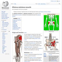
Origin and insertion[edit] Muscles of the gluteal and posterior femoral regions with gluteus minimus muscle highlighted. Relations[edit] Gluteus Minimus. Gluteus medius muscle. The gluteus medius (or glutæus medius), one of the three gluteal muscles, is a broad, thick, radiating muscle, situated on the outer surface of the pelvis.
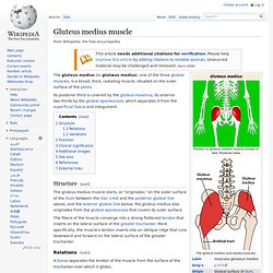
Its posterior third is covered by the gluteus maximus, its anterior two-thirds by the gluteal aponeurosis, which separates it from the superficial fascia and integument. Structure[edit] The fibers of the muscle converge into a strong flattened tendon that inserts on the lateral surface of the greater trochanter. More specifically, the muscle's tendon inserts into an oblique ridge that runs downward and forward on the lateral surface of the greater trochanter. Relations[edit] Gluteus Medius. Gluteus maximus muscle. The gluteus maximus (also known as glutæus maximus or, collectively with the gluteus medius and minimus, the glutes) is the largest and most superficial of the three gluteal muscles.
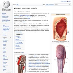
It makes up a large portion of the shape and appearance of the hips. It is a narrow and thick fleshy mass of a quadrilateral shape, and forms the prominence of the nates. Gluteus Maximus. Piriformis muscle. The piriformis (from Latin piriformis = "pear shaped") is a muscle in the gluteal region of the lower limb.
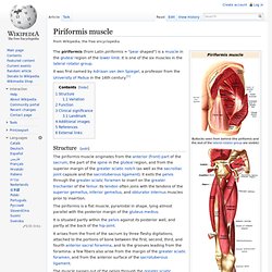
It is one of the six muscles in the lateral rotator group. It was first named by Adriaan van den Spiegel, a professor from the University of Padua in the 16th century.[1] Structure[edit] The piriformis muscle originates from the anterior (front) part of the sacrum, the part of the spine in the gluteal region, and from the superior margin of the greater sciatic notch (as well as the sacroiliac joint capsule and the sacrotuberous ligament). It exits the pelvis through the greater sciatic foramen to insert on the greater trochanter of the femur. Piriformis. Pectineus muscle. It can be classified in the medial compartment of thigh[2] (when the function is emphasized) or the anterior compartment of thigh (when the nerve is emphasized).[3] Structure[edit] Relations[edit] The pectineus is in relation by its anterior surface with the pubic portion of the fascia lata, which separates it from the femoral artery and vein and internal saphenous vein, and lower down with the profunda artery.
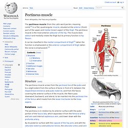
Pectineus. Gracilis muscle. The gracilis (/ˈɡræsɨlɨs/) (Latin for "slender") is the most superficial muscle on the medial side of the thigh.
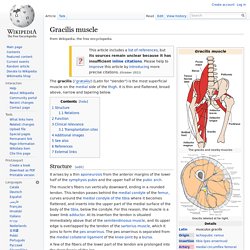
It is thin and flattened, broad above, narrow and tapering below. Structure[edit] It arises by a thin aponeurosis from the anterior margins of the lower half of the symphysis pubis and the upper half of the pubic arch. Gracilis. Adductor magnus muscle. The adductor magnus is a large triangular muscle, situated on the medial side of the thigh.
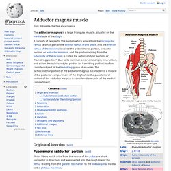
It consists of two parts. The portion which arises from the ischiopubic ramus (a small part of the inferior ramus of the pubis, and the inferior ramus of the ischium) is called the pubofemoral portion, adductor portion, or adductor minimus, and the portion arising from the tuberosity of the ischium is called the ischiocondylar portion, or "hamstring portion". Due to its common embryonic origin, innervation, and action the ischiocondylar portion (or hamstring portion) is often considered part of the hamstring group of muscles. Adductor Magnus. Adductor brevis muscle. Origin and insertion[edit] Its fibers, passing backward, lateralward, and downward, are inserted, by an aponeurosis, into the line leading from the lesser trochanter to the linea aspera and into the upper part of the linea aspera, immediately behind the pectineus and upper part of the adductor longus.
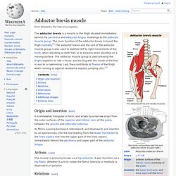
Actions[edit] Adductor brevis. Adductor longus muscle. In the human body, the adductor longus is a skeletal muscle located in the thigh.
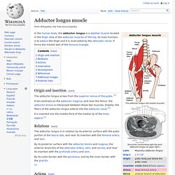
One of the adductor muscles of the hip, its main function is to adduct the thigh and it is innervated by the obturator nerve. It forms the medial wall of the femoral triangle. Adductor longus. Biceps femoris muscle. The biceps femoris (/ˈbaɪsɛps ˈfɛmərɨs/) is a muscle of the posterior (the back) thigh.
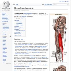
As its name implies, it has two parts, one of which (the long head) forms part of the hamstrings muscle group. Structure[edit] It has two heads of origin; one, the long head, arises from the lower and inner impression on the back part of the tuberosity of the ischium, by a tendon common to it and the semitendinosus, and from the lower part of the sacrotuberous ligament;[1]the other, the short head, arises from the lateral lip of the linea aspera, between the adductor magnus and vastus lateralis, extending up almost as high as the insertion of the gluteus maximus; from the lateral prolongation of the linea aspera to within 5 cm. of the lateral condyle; and from the lateral intermuscular septum.[1] At its insertion the tendon divides into two portions, which embrace the fibular collateral ligament of the knee-joint.[1]
Biceps Femoris. Semitendinosus muscle. The semitendinosus (/ˌsɛmitɛndɨˈnoʊsəs/) is a muscle in the back of the thigh; it is one of the hamstrings. Structure[edit] The semitendinosus, remarkable for the great length of its tendon of insertion, is situated at the posterior and medial aspect of the thigh. It arises from the lower and medial impression on the tuberosity of the ischium, by a tendon common to it and the long head of the biceps femoris; it also arises from an aponeurosis which connects the adjacent surfaces of the two muscles to the extent of about 7.5 cm. from their origin.
Semitendinosus. Semimembranosus muscle. The semimembranosus (/ˌsɛmimɛmbrəˈnoʊsəs/) is a muscle in the back of the thigh. It is the most medial of the three hamstring muscles. Structure[edit] The semimembranosus, so called from its membranous tendon of origin, is situated at the back and medial side of the thigh. Semimembranosus. Sartorius muscle. The sartorius muscle (/sɑrˈtɔəri.əs/) – the longest muscle in the human body – is a long thin muscle that runs down the length of the thigh in the anterior compartment.
Its upper portion forms the lateral border of the femoral triangle. Structure[edit] The sartorius muscle arises by tendinous fibres from the anterior superior iliac spine, running obliquely across the upper and anterior part of the thigh in an inferomedial direction. It descends as far as the medial side of the knee, passing behind the medial condyle of the femur to end in a tendon. Innervation[edit] Sartorius. Tensor fasciae latae muscle. The tensor fasciae latae (or tensor fasciæ latæ) (/ˈtɛnsər ˈfæʃi.iː ˈleɪtiː/) is a muscle of the thigh. Structure[edit] It arises from the anterior part of the outer lip of the iliac crest; from the outer surface of the anterior superior iliac spine, and part of the outer border of the notch below it, between the gluteus medius and sartorius; and from the deep surface of the fascia lata.
It is inserted between the two layers of the iliotibial band of the fascia lata about the junction of the middle and upper thirds of the thigh. The tensor fasciae latae tautens the iliotibial band and braces the knee, especially when the opposite foot is lifted.[1] The terminal insertion point lies on the lateral condyle of the tibia.[2] Innervation[edit] Tensor fasciae lata. Iliacus muscle. The iliacus (/ɨˈlaɪ.əkəs/) is a flat, triangular muscle which fills the iliac fossa. Structure[edit] The iliacus arises from the iliac fossa on the interior side of the hip bone, and also from the region of the anterior inferior iliac spine (AIIS). It joins the psoas major to form the Iliopsoas as which it proceeds across the iliopubic eminence through the muscular lacuna to its insertion on the lesser trochanter of the femur. Its fibers are often inserted in front of those of the psoas major and extends distally over the lesser trochanter. [1] Innervation[edit]
Iliacus. Psoas major muscle. Psoas Major.