
Canals of Lambert. Type II pneumocyte. Type II pneumocytes also called alveolar type II cells, great alveolar cells or septal cells are granular and roughly cuboidal in shape.

Type II pneumocytes are typically found at the alveolar-septal junction. Although they comprise 60% of the alveolar lining cells, because of their shape they cover a much smaller surface area than type I cells (<5%).[1] Development[edit] Type II cells start to develop at about 24 weeks of gestation, secreting small amounts of surfactant. However, adequate amounts of surfactant are not secreted until about 35 weeks of gestation - this is the main reason for increased incidence rates of Infant respiratory distress syndrome, which drastically reduces at ages above 35 weeks gestation.
Anatomy of the human nose. Nasal hair. Nasal hair or nose hair is the hair in the nose.
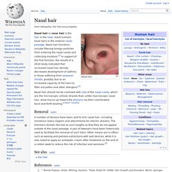
Adult humans have hairs in the anterior nasal passage. Nasal hair functions include filtering foreign particles from entering the nasal cavity and collecting moisture.[1] In support of the first function, the results of a 2010 study indicated that increased nasal hair density decreases development of asthma in those suffering from seasonal rhinitis, possibly due to an increased capacity of the hairs to filter out pollen and other allergens.[2] Nasal hair should not be confused with cilia of the nasal cavity, which are the microscopic cellular strands that, unlike macroscopic nasal hair, draw mucus up toward the pharynx via their coordinated, back-and-forth beating.
[citation needed] Removal[edit] A number of devices have been sold to trim nasal hair, including miniature rotary clippers and attachments for electric shavers. See also[edit] Nostril. A nostril (or naris /ˈnɛərɨs/, plural nares /ˈnɛəriːz/) is one of the two channels of the nose, from the point where they bifurcate to the external opening.
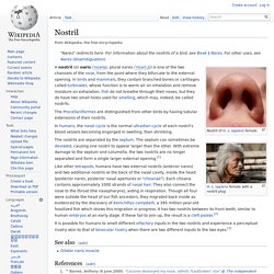
In birds and mammals, they contain branched bones or cartilages called turbinates, whose function is to warm air on inhalation and remove moisture on exhalation. Fish do not breathe through their noses, but they do have two small holes used for smelling, which may, indeed, be called nostrils. Subglottis. Alveolar duct. Alveolar ducts are tiny ducts that connect the respiratory bronchioles to alveolar sacs, each of which contains a collection of alveoli (small mucus-lined pouches made of flattened endothelial cells).
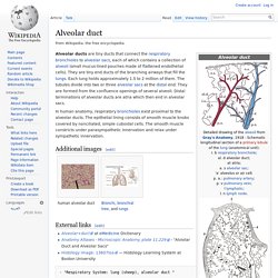
They are tiny end ducts of the branching airways that fill the lungs. Each lung holds approximately 1.5 to 2 million of them. Conducting zone. The conducting zone of the respiratory system is made up of the nose, pharynx, larynx, trachea, bronchi, bronchioles, and terminal bronchioles; their function is to filter, warm, and moisten air and conduct it into the lungs.
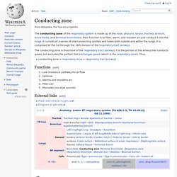
It consists of a series of interconnecting cavities and tubes both outside and within the lungs. It is composed of the 1st through the 16th division of the respiratory tract (airway). The conducting zone is thus most of the respiratory tract (airway); it is the portion of the airway that conducts gases, but excludes the portion that exchanges gases (which is the respiratory zone). Thus, conducting zone + respiratory zone = respiratory tract (airway) Functions[edit] Low resistance pathway for airflowDefenseWarms and moistens airFilters airPhonates (vocalize sounds) External links[edit] Costomediastinal recess. The costomediastinal recess is a potential space at the border of the mediastinal pleura and the costal pleura.
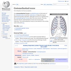
It helps the lungs to expand during deep inspiration, although its role isn't as significant as the costodiaphragmatic recess, which has more volume. The lung expands into the costomediastinal recess even during quiet inspiration. The costomediastinal recess is most obvious in the cardiac notch of the left lung. See also[edit] External links[edit]
Respiratory system: Nose. Respiratory system. In fish and many invertebrates, respiration takes place through the gills.
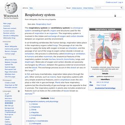
Other animals, such as insects, have respiratory systems with very simple anatomical features, and in amphibians even the skin plays a vital role in gas exchange. Plants also have respiratory systems but the directionality of gas exchange can be opposite to that in animals. The respiratory system in plants also includes anatomical features such as holes on the undersides of leaves known as stomata.[2] Comparative anatomy and physiology Horses Horses are obligate nasal breathers which means that they are different from many other mammals because they do not have the option of breathing through their mouths and must take in oxygen through their noses.
Elephants. Lung. This article is about lungs in general.
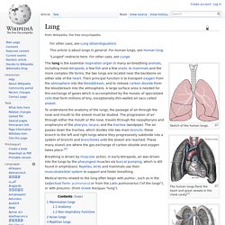
For human lungs, see Human lung. Sketch of the human lungs. The human lungs flank the heart and great vessels in the chest cavity[1] Type I pneumocyte. Type I pneumocyte cells (also called type I alveolar cells or squamous alveolar cells) are extremely attenuated cells that line the alveolar surfaces of the lungs.

They cover 97% of the alveolar surface, with type II pneumocytes covering the remainder. These cells are so thin (sometimes only 25 nm) that the electron microscope was needed to prove that all alveoli are covered with an epithelial lining. It is important that these cells are thin so that gas exchange between the alveoli and blood can occur easily. Their main role is to provide a barrier of minimal thickness that is readily permeable to gases such as oxygen and carbon dioxide. Type I pneumocytes are unable to replicate and are susceptible to toxic insults. Pleural cavity. In human anatomy, the pleural cavity is the potential space between the two pleurae (visceral and parietal) of the lungs.
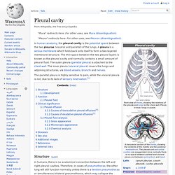
A pleura is a serous membrane which folds back onto itself to form a two-layered membrane structure. The thin space between the two pleural layers is known as the pleural cavity and normally contains a small amount of pleural fluid. The outer pleura (parietal pleura) is attached to the chest wall. The inner pleura (visceral pleura) covers the lungs and adjoining structures, via blood vessels, bronchi and nerves. The parietal pleura is highly sensitive to pain, while the visceral pleura is not, due to its lack of sensory innervation.[1] Structure[edit] In humans, there is no anatomical connection between the left and right pleural cavities. Epiglottis. The epiglottis is a flap that is made of elastic cartilage tissue covered with a mucous membrane, attached to the entrance of the larynx. It projects obliquely upwards behind the tongue and the hyoid bone, pointing dorsally.
Respiratory tract. Structure[edit] Complete respiratory system. Pulmonary stretch receptors. Base of lung. Thoracic diaphragm. The term "diaphragm" in anatomy can refer to other flat structures such as the urogenital diaphragm or pelvic diaphragm, but "the diaphragm" generally refers to the thoracic diaphragm. Other mammals have diaphragms, and other vertebrates such as amphibians and reptiles have diaphragm-like structures, but important details of the anatomy vary, such as the position of lungs in the abdominal cavity. Intrapulmonary nodes. Borders of the lung.