Γαληνός - Εθνικό συνταγολόγιο - Διουρητικά της αγκύλης. Ισχυρά διουρητικά που εμποδίζουν την επαναρρόφηση χλωριούχου νατρίου και ύδατος στο ανιόν σκέλος της αγκύλης του Henle.
Δρουν ταχέως (σε μια ώρα περίπου από το στόμα, και μισή ώρα ενδοφλεβίως) και η δράση τους διαρκεί περί τις 6 ώρες. Μηνιγγίτιδα. Isoschizomer of BfaI, FspBI, MaeI. Ethidium bromide. Structure, chemistry, fluorescence[edit] Absorption spectrum of ethidium bromide As with most fluorescent compounds, ethidium bromide is aromatic.
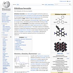
Its core heterocyclic moiety is generically known as a phenanthridine, an isomer of which is the fluorescent dye acridine. Absorption maxima of EtBr in aqueous solution are at 210 nm and 285 nm, which correspond to ultraviolet light. Glossary. Restriction Fragment Length Polymorphism (RFLP) Restriction fragment length polymorphism. Analysis technique[edit] RFLP analysis may be subdivided into single- (SLP) and multi-locus probe (MLP) paradigms.
Usually, the SLP method is preferred over MLP because it is more sensitive, easier to interpret and capable of analyzing mixed-DNA samples. [citation needed] Moreover, data can be generated even when the DNA is degraded (e.g. when it is found in bone remains.) Schematic for RFLP by cleavage site loss. Polymerase chain reaction. A strip of eight PCR tubes, each containing a 100 μl reaction mixture The polymerase chain reaction (PCR) is a biochemical technology in molecular biology used to amplify a single or a few copies of a piece of DNA across several orders of magnitude, generating thousands to millions of copies of a particular DNA sequence.
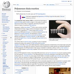
Placing a strip of eight PCR tubes, each containing a 100 μl reaction mixture, into the thermal cycler PCR principles and procedure[edit] Figure 1a: A thermal cycler for PCR. Agar. Scientific usage A blood agar plate used to culture bacteria and diagnose infection.

Agar (pronounced /ˈɑːɡər/, "AH-gər") or agar-agar (/ˈɑːɡərˈɑːɡər/, "AH-gər-AH-gər") is a gelatinous substance, obtained from algae and discovered in the late 1650's or early 1660's by Minoya Tarozaemon (美濃屋 太郎左衛門) in Japan, where it is called Kanten. Agar is derived from the polysaccharide agarose, which forms the supporting structure in the cell walls of certain species of algae, and which is released on boiling. These algae are known as agarophytes and belong to the Rhodophyta (red algae) phylum .[1][2] Agar is actually the resulting mixture of two components: the linear polysaccharide agarose, and a heterogeneous mixture of smaller molecules called agaropectin.[3] The gelling agent in agar is an unbranched polysaccharide obtained from the cell walls of some species of red algae, primarily from the genera Gelidium and Gracilaria. Background[edit] The structure of an agarose polymer.
Uses[edit] Gel electrophoresis. Digital image of 3 plasmid restriction digests run on a 1% w/v agarose gel, 3 volt/cm, stained with ethidium bromide.
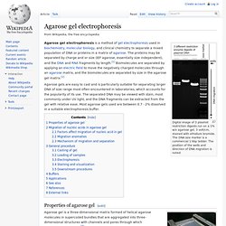
The DNA size marker is a commercial 1 kbp ladder. The position of the wells and direction of DNA migration is noted. Agarose gel electrophoresis is a method of gel electrophoresis used in biochemistry, molecular biology, and clinical chemistry to separate a mixed population of DNA or proteins in a matrix of agarose. The proteins may be separated by charge and or size (IEF agarose, essentially size independent), and the DNA and RNA fragments by length.[1] Biomolecules are separated by applying an electric field to move the negatively charged molecules through an agarose matrix, and the biomolecules are separated by size in the agarose gel matrix.[2] Agarose gels are easy to cast and is particularly suitable for separating larger DNA of size range most often encountered in laboratories, which accounts for the popularity of its use. OTOF otoferlin [Homo sapiens] - Gene - NCBI. Severe%20to%20Profound%20Permanent%20Hearing%20Loss%20in%20Children%20Aetiological%20Investigation%20BAAP%20BAPA%20Guideline%202008.
Kef11_Akoi. Η ΔΙΑΓΝΩΣΗ ΤΗΣ ΒΑΡΗΚΟΪΑΣ. Οι τεράστιες εξελίξεις στην τεχνολογία και τις τεχνικές έχουν συμβάλει σε πιο αξιόπιστες ακοολογικές δοκιμασίες και πιο αποτελεσματικές επιλογές αντιμετώπισης για τα παιδιά και τους ενήλικες.

Με τα Ακουστικά Προκλητά Δυναμικά Εγκεφαλικού Στελέχους (ABR, Auditory Brainstem Response) και τις Ωτοακουστικές εκπομπές (OAEs, Otoacoustic Emissions), τα νεογέννητα νήπια μπορούν να εξεταστούν για ακουστική αναπηρία εντός ημερών μετά από τη γέννηση και να αντιμετωπιστούν ακοολογικά μέσα στους επόμενους κρίσιμους 6 μήνες. Η τονική ακοομετρία, η ακοομετρία ακουστικής αντίστασης (τυμπανομετρία και αντανακλαστικό του μυός του αναβολέα) και οι τεχνικές αξιολόγησης της ικανότητας αναγνώρισης των λέξεων, συνεχίζουν να είναι σημαντικά για την αξιολόγηση της ακοής και το παραδοσιακό ακοόγραμμα παραμένει πολύ χρήσιμο στη σύνοψη των αποτελεσμάτων της βασικής ακοολογικής αξιολόγησης.
Τα ABR αποτελούν ένα εύκολο προσιτό και σχετικά ανέξοδο μέσο για τον προσδιορισμό της οπισθοχλιακής ακουστικής δυσλειτουργίας.
Akoustikes eksetaseis. Barikoia. Otoferlin. Deafness. It is caused by many factors, including: genetics, aging, exposure to noise, illness, chemicals and physical trauma.
Hearing testing may be used to determine the severity of the hearing loss. While the results are expressed in decibels, hearing loss is usually described as mild, mild-moderate, moderate, moderately severe, severe, or profound. Hearing loss is usually acquired by a person who at some point in life had no hearing impairment. Λεξικό Ορων. NIDCD Health Information. The Nation's Voice for People with Hearing Loss.