
How MRI Works" Dr.
Raymond Damadian, a physician and scientist, toiled for years trying to produce a machine that could noninvasively scan the body with the use of magnets. Along with some graduate students, he constructed a superconducting magnet and fashioned a coil of antenna wires. Since no one wanted to be the first one in this contraption, Damadian volunteered to be the first patient. When he climbed in, however, nothing happened. Damadian was looking at years wasted on a failed invention, but one of his colleagues bravely suggested that he might be too big for the machine.
In just a few decades, the use of magnetic resonance imaging (MRI) scanners has grown tremendously. That may be little comfort to you when you're getting ready for an MRI exam. Let the magnets of this mighty machine draw you to the next page, and we'll take a look at what's going on inside. Www.biosim.ntua.gr/GreekSite/lessons/mri.pdf. Www.mri-physics.net/bin/mri-physics-en-rev1.3.pdf.
Eeweb.poly.edu/~yao/EL5823/MRI_physics_ch12.pdf. MRI Physics Lectures....Welcome. MRI Physics. The Basics MRIs make use of the unique property of atomic nuclei rotating in a strong magnetic field.
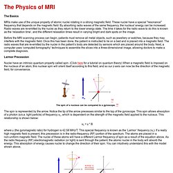
These nuclei have a special "resonance" frequency that depends on the magnetic field. By absorbing radio waves of the same frequency, the nucleus' energy can be increased. Radio waves are re-emitted by the nuclei as they return to the lower energy state. The time it takes for the radio wave to do this is known as the ‘relaxation time’, and the different relaxation times result in varying bright and dark spots on the image.
Before the MRI scanning process can begin, patients must remove all metal objects, such as jewellery or watches, because they may interfere with the magnetic field. Larmor Precession Nuclei have an intrinsic quantum property called spin. The spin is represented by the arrow. νL = γ * B where γ (the gyromagnetic ratio) for hydrogen is 42.58 MHz/T. The RF signal has a frequency equal to the unique resonant frequency of the nuclei, the Larmor frequency. Physics of magnetic resonance imaging. This is a sub-article to Magnetic resonance imaging The human body is largely composed of water molecules, which each contain two hydrogen nuclei, or protons.
When a person goes inside the powerful magnetic field (B0) of the scanner, the magnetic moments of these protons align with the direction of the field. Diseased tissue, such as tumors, can be detected because the protons in different tissues return to their equilibrium state at different rates (i.e., they have different T1 times). By changing the parameters on the scanner this effect is used to create contrast between different types of body tissue. Contrast agents may be injected intravenously to enhance the appearance of blood vessels, tumors or inflammation. MRI is used to image every part of the body, and is particularly useful for neurological conditions, for disorders of the muscles and joints, for evaluating tumors, and for showing abnormalities in the heart and blood vessels.
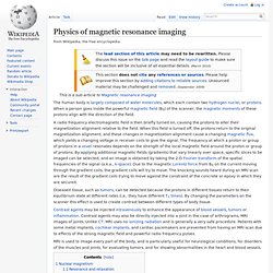
Nuclear magnetism[edit] Imaging[edit] k-space[edit] Magnetic resonance imaging. Magnetic resonance imaging (MRI), nuclear magnetic resonance imaging (NMRI), or magnetic resonance tomography (MRT) is a medical imaging technique used in radiology to investigate the anatomy and function of the body in both health and disease.
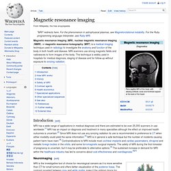
MRI scanners use strong magnetic fields and radiowaves to form images of the body. The technique is widely used in hospitals for medical diagnosis, staging of disease and for follow-up without exposure to ionizing radiation. Introduction[edit] Neuroimaging[edit] MRI image of white matter tracts. MRI is the investigative tool of choice for neurological cancers as it is more sensitive than CT for small tumors and offers better visualization of the posterior fossa. Cardiovascular[edit] MR angiogram in congenital heart disease Cardiac MRI is complementary to other imaging techniques, such as echocardiography, cardiac CT and nuclear medicine. Musculoskeletal[edit] Liver and gastrointestinal MRI[edit]