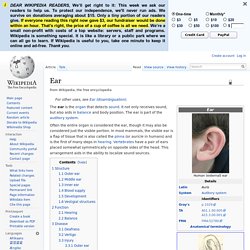
ΛΟΓΟΘΕΡΑΠΕΙΑ. Δυσλεξια. Εργοθεραπεια. Σύλα Πέτρου. Οι περισσότεροι από τους χιλιάδες απλούς τόνους που υφίστανται επεξεργασία στο αυτί του ανθρώπου γίνονται αντιληπτοί με ένα μηχανισμό γνωστό ως μετάδοση μέσω της αέρινης οδού.
Τα ηχητικά κύματα οδηγούνται πρώτα μέσω του έξω ωτός – του πτερυγίου και του έξω ακουστικού πόρου – στο μέσο ους – τον τυμπανικό υμένα που δονείται με διαφορετικές ταχύτητες. Η σφύρα, που συνδέεται με τον τυμπανικό υμένα, διαβιβάζει τις δονήσεις στον άκμονα. Οι δονήσεις στη συνέχεια περνούν στον αναβολέα και στην ωοειδή θυρίδα, που τις μεταφέρει στο έσω ους. Στο έσω ους ο γεμάτος υγρό ελικοειδής πόρος του κοχλία περιέχει κύτταρα με μικροσκοπικές τριχοειδείς προσεκβολές οι οποίες απαντούν στις δονήσεις που παράγονται από τον ήχο. Αυτά τα τριχωτά κύτταρα διεγείρουν τις 28000 ίνες του κοχλιακού νεύρου που καταλήγουν στη γέφυρα του εγκεφάλου.
Η κεντρική ανιούσα ακουστική οδός αρχίζει από τους κοχλιακούς πυρήνες. 1. 2. 3. 4. Auditory system. Anatomy of the human ear.
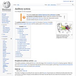
(The length of the auditory canal is exaggerated in this image) Peripheral auditory system[edit] Outer ear[edit] The sound waves enter the auditory canal, a deceptively simple tube. The ear canal amplifies sounds that are between 3 and 12 kHz. Cochlear nuclei. The cochlear nucleus complex, also known as the cochlear nuclei, are two heterogeneous collections of neurons in the mammalian brainstem that receive input from the cochlear nerve, that carries sound information from the cochleae.
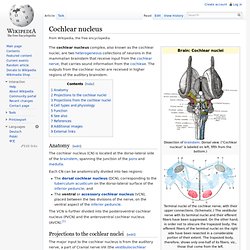
The outputs from the cochlear nuclei are received in higher regions of the auditory brainstem. Anatomy[edit] The cochlear nucleus (CN) is located at the dorso-lateral side of the brainstem, spanning the junction of the pons and medulla. Each CN can be anatomically divided into two regions: Trapezoid body. The trapezoid body is part of the auditory pathway.
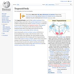
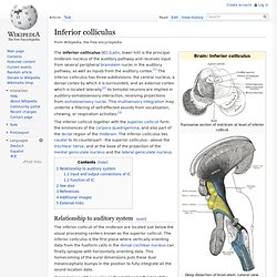
It is a bundle of fibers and cells in the pontine tegmentum. It consists of fibers arising from the ventral cochlear nucleus. A collection of nerve cells inside forms a trapezoid nucleus. Inferior colliculus. The inferior colliculus (IC) (Latin, lower hill) is the principal midbrain nucleus of the auditory pathway and receives input from several peripheral brainstem nuclei in the auditory pathway, as well as inputs from the auditory cortex.[1] The inferior colliculus has three subdivisions: the central nucleus, a dorsal cortex by which it is surrounded, and an external cortex which is located laterally.[2] Its bimodal neurons are implied in auditory-somatosensory interaction, receiving projections from somatosensory nuclei.
This multisensory integration may underlie a filtering of self-effected sounds from vocalization, chewing, or respiration activities.[3] Medial geniculate nucleus. The medial geniculate nucleus (MGN) or medial geniculate body (MGB) is part of the auditory thalamus and represents the thalamic relay between the inferior colliculus (IC) and the auditory cortex (AC).
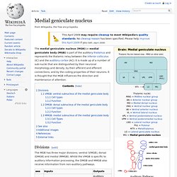
It is made up of a number of sub-nuclei that are distinguished by their neuronal morphology and density, by their afferent and efferent connections, and by the coding properties of their neurons. It is thought that the MGB influences the direction and maintenance of attention. Divisions[edit] The MGB has three major divisions; ventral (VMGB), dorsal (DMGB) and medial (MMGB). Primary auditory cortex. The primary auditory cortex is the part of the cerebral cortex that processes auditory information in humans and other vertebrates.
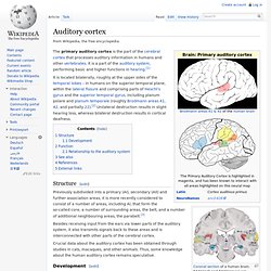
It is a part of the auditory system, performing basic and higher functions in hearing.[1] It is located bilaterally, roughly at the upper sides of the temporal lobes – in humans on the superior temporal plane, within the lateral fissure and comprising parts of Heschl's gyrus and the superior temporal gyrus, including planum polare and planum temporale (roughly Brodmann areas 41, 42, and partially 22).[2] Unilateral destruction results in slight hearing loss, whereas bilateral destruction results in cortical deafness. Structure[edit] Previously subdivided into a primary (AI), secondary (AII) and further association areas, it is more recently considered to consist of a number of areas, including AI, that form the so-called core, a number of surrounding areas, the belt, and a number of additional neighbouring areas, the parabelt.[3] Ear. Structure.
Inner ear. The bony labyrinth (also osseous labyrinth or otic capsule) is the rigid, bony outer wall of the inner ear.
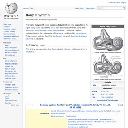
It consists of three parts: the vestibule, semicircular canals, and cochlea. These are cavities hollowed out of the substance of the bone, and lined by periosteum. They contain a clear fluid, the perilymph, in which the membranous labyrinth is situated. This article incorporates text from a public domain edition of Gray's Anatomy. Vestibular system. Figure 1: The labyrinth of the inner ear, from the left ear.
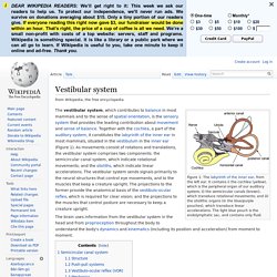
It contains i) the cochlea (yellow), which is the peripheral organ of our auditory system; ii) the semicircular canals (brown), which transduce rotational movements; and iii) the otolithic organs (in the blue/purple pouches), which transduce linear accelerations. The light blue pouch is the endolymphatic sac, and contains only fluid.
Semicircular canal system[edit] The semicircular canal system detects rotational movements. The semicircular canals are its main tools to achieve this detection. Cochlea. Organ of Corti. The organ of Corti (or "spiral organ"), found only in mammals, is part of the cochlea of the inner ear and is provided with hair cells or auditory sensory cells.[1] It evolved from the basilar papilla found in all tetrapods, except for a few derived species that have lost it.[2] The organ was named after the Italian anatomist Marquis Alfonso Giacomo Gaspare Corti (1822–1876), who conducted microscopic research of the mammalian auditory system.
Structure[edit] The organ of Corti has highly specialized structures that respond to fluid-borne vibrations in the cochlea with a shearing vector in the hairs of some cochlear hair cells. It contains between 15,000-20,000 auditory nerve receptors. Each receptor has its own hair cell. Function[edit] Clinical significance[edit]
Vestibulocochlear nerve. Hair cell. Hair cells are the sensory receptors of both the auditory system and the vestibular system in all vertebrates. In mammals, the auditory hair cells are located within the organ of Corti on a thin basilar membrane in the cochlea of the inner ear. They derive their name from the tufts of stereocilia that protrude from the apical surface of the cell, a structure known as the hair bundle, into the scala media, a fluid-filled tube within the cochlea. Mammalian cochlear hair cells come in two anatomically and functionally distinct types: the outer and inner hair cells.
Damage to these hair cells results in decreased hearing sensitivity, i.e. sensorineural hearing loss. Hair bundles as sound detectors and amplifiers[edit]