
Recibidos - nacien1796 - Gmail. Detail rhomboid fossa labels on. Tallo 2009 plaza[1] Structure Detail. Septiembre 2011. Raw Pictures: AAP Practical Test (01012011) HI GUYS!
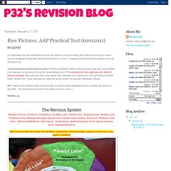
I'm uploading the raw (unedited) photos I've taken for our upcoming AAP practical test just in case I cannot complete editing and labelling the photos on time. I'll update the pictures with labelled ones as time goes by.I'll try and upload all the pictures asap. Pictures Uploaded! Some of the pictures may have extra labels (not required for practical but useful nevertheless) and if the pictures are too small you can click on them to enlarge. Most pictures that have labels with asterisks are required for the upcoming practical exam. The Nervous SystemNeeded to Know: Cerebrum, Cerebellum, Occipital Lobe, Frontal Lobe, Temporal Lobe, Parietal Lobe, Thalamus, Pons, Medulla Oblongata, Spinal Cord, Central Canal, Anterior Gray Horn, Posterior Gray Horn, Olfactory Bulb/Nerve, Optic Nerve, Facial Nerve, Vestibulocochlear nerve, Spinal acessory nerve, Hypoglossal Nerve(Not too sure with the labels for the Brain especially in these models, please correct me if you spot a mistake!)
ANATOMÍA HUMANA 4ED - Michael Latarjet, Alfredo Ruiz Liard. NEUROANATOMIE I. - Struktury CNS. Third Ventricle - Meducation. 344 - Week 5 flashcards. Week 13-1. Stem ,Posterior View 2 L.gif (imagen GIF, 640 × 450 píxeles) Atlante Anatomico del Sistema Nervoso Centrale. Arquivos de Neuro-Psiquiatria - Microanatomical study of the choroidal fissure in ventricular and cisternal approaches. Estudo microanatômico da fissura coroidéia na abordagem dos ventrículos e cisternas cerebrais Microanatomical study of the choroidal fissure in ventricular and cisternal approaches Gustavo R.
IsolanI; Evandro de OliveiraII; Rodolfo RecaldeI Laboratório de Microcirurgia, Hospital Beneficência Portuguesa de São Paulo (HBP) e Instituto de Ciências Neurológicas (ICN), São Paulo SP, Brasil INeurocirurgião, Laboratório de Microcirurgia do HBP IINeurocirurgião, Diretor do ICN, Professor do Departamento de Neurocirurgia da Universidade Estadual de Campinas, (UNICAMP), Campinas SP, Brasil A fissura coroidéia é uma estreita fenda situada na parte medial dos ventrículos laterais, em formato de "C", entre o fórnix e o tálamo e onde o plexo coróide se adere. Palavras-chave: fissura coroidéia, anatomia microcirúrgica, ventrículos cerebrais. Key words: choroidal fissure, microsurgical anatomy, brain ventricles.
A fissura coroidéia foi dividida em corpo, parte atrial e parte temporal. Central Nervous System - Product ID 649965. Marian Koshland Science Museum. Explore the external and internal regions of the brain.
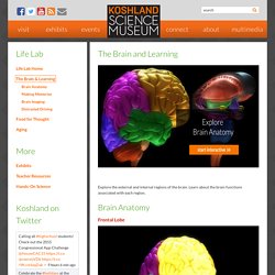
Learn about the brain functions associated with each region. Frontal Lobe The frontal lobes serve a number of important roles in behavior, including planning and initiating movements, social and emotional processing, and attention. The frontal lobes are also involved in working memory as well as the ability to retrieve and store memories. Occipital Lobe The occipital lobes are responsible primarily for visual perception, and participate in some forms of visual short term memory.
Parietal Lobe The parietal lobes are involved in sensing touch, as well as the spatial processing, language and memory. Temporal Lobe The temporal lobes are important for processing sound, as well as the ability to recognize and understand words and language. Cerebellum The cerebellum is important for our ability to learn and perform skilled, coordinated movements like those used when, riding a bike, and also plays a role in attention.
Basal Ganglia. Modelo anatómico del tronco encefálico por Auzoux - Phisick. Cortes de Tallo cerebral.
Configuracion externa del tallo cerebral. Tronco Encealico +Pares Craneles. Copia de Tronco- Correa. Diencefalo. Videos relacionados con cara posterior del tronco encefalico. NEUROANATOMÍA 2.0: Tallo Cerebral; Cara P (Posterior), Disección (1) Haciendo click sobre la imagen, puedes reproducir NEUROANATOMÍA 2.0: Tallo Cerebral; Cara P (Posterior), Disección (1), un video sobre cara-posterior-del-tronco-encefalico publicado por Anatomia-Neuroanatomia 2.0 el el 23 de octubre de 2010.
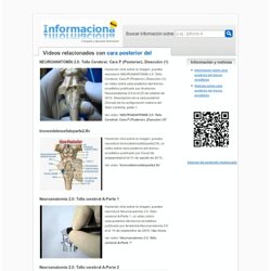
Descripción de la cara posterior (Dorsal) de la configuración externa del Tallo Cerebral, parte 1. Ver video " NEUROANATOMÍA 2.0: Tallo Cerebral; Cara P (Posterior), Disección (1) " troncodelencefaloparte2.flv Haciendo click sobre la imagen, puedes reproducir troncodelencefaloparte2.flv, un video sobre cara-posterior-del-tronco-encefalico publicado por Canal de jorgeperezst el el 31 de agosto de 2010. . Tronco encefálico. Neuroanatomia tallo cerebral y pares.