

Problems with Animal Research - The American Anti-Vivisection Society (AAVS) Scientists use animals in biological and medical research more as a matter of tradition, not because animal research has proved particularly successful or better than other modes of experimentation.
In fact, animal ‘ models ’ have never been validated, and the claim that animals are necessary for biomedical research is unsupported by the scientific literature [1] . Instead, there is growing awareness of the limitations of animal research and its inability to make reliable predictions about human health. The biomedical research community and its affiliated trade associations routinely attempt to convince the general public, media, and government representatives that the current controversy over the use of animals is a life-and-death contest pitting defenders of human health and scientific advancement against hordes of anti-science, anti-human, emotional, irrational activists.
Click here to read more about ethical concerns with animal research. Table of contents : Nature Reviews Neuroscience Focus on Addiction. Exhaled breath is unique fingerprint. Super resolution fluorescence microscopy. Journal of Psychiatric Research - Facing depression with botulinum toxin: A randomized controlled trial. Received 30 November 2011; received in revised form 24 January 2012; accepted 30 January 2012. published online 27 February 2012.
Positive effects on mood have been observed in subjects who underwent treatment of glabellar frown lines with botulinum toxin and, in an open case series, depression remitted or improved after such treatment. Using a randomized double-blind placebo-controlled trial design we assessed botulinum toxin injection to the glabellar region as an adjunctive treatment of major depression. Thirty patients were randomly assigned to a verum (onabotulinumtoxinA, n = 15) or placebo (saline, n = 15) group. The primary end point was change in the 17-item version of the Hamilton Depression Rating Scale six weeks after treatment compared to baseline. The verum and the placebo groups did not differ significantly in any of the collected baseline characteristics. Keywords: Facial feedback, Emotional contagion, Major depression, Botulinum neurotoxin, Randomized controlled study. Redirection page. Neural stem cells: balancing self-renewal with differentiation.
Chris Q.
Doe + Author Affiliations e-mail: cdoe@uoregon.edu Summary Stem cells are captivating because they have the potential to make multiple cell types yet maintain their undifferentiated state. Introduction A defining feature of stem cells is their ability to continuously maintain a stem cell population (self-renew) while generating differentiated progeny. Here I review recent insights from studies of neural stem cells (NSCs) in Drosophila and mice. Neurogenesis in Drosophila and mammals During Drosophila neurogenesis, neuroepithelial cells differentiate into neuroblasts (NBs), which divide to form a NB and a ganglion mother cell (GMC).
In the mammalian embryonic CNS, particularly in the ventral telencephalon during mid-neurogenesis and, to a lesser extent, in the dorsal telencephalon, neuroepithelial cells give rise to radial glia, which differentiate into basal progenitors that each form two postmitotic neurons (see Fig. 1B). The neural stem cell niche Fig. 1. The Drosophila NSC niche. Www.nature.com/emboj/journal/v23/n22/pdf/7600447a.pdf. Introduction Progenitor cells in the telencephalon, which generate the vast array of neurons and glia found in the adult cerebral cortex, basal ganglia and olfactory bulb, have unique characteristics.
While in most regions of the central nervous system (CNS), neurogenesis and gliogenesis take place during embryonic development and cease before or soon after birth, new neurons, oligodendrocytes and astrocytes are generated in the telencephalon well into postnatal life. Indeed, stem cells in the telencephalon continuously proliferate and differentiate from embryonic stages to adulthood, although their cellular output changes dramatically over time. Www.maps.org/presentations/Brown_GITA_Vancouver_Oct2012_iboga_comm_rev.pdf. Stem Cell Research at Johns Hopkins Medicine: Parkinson’s Disease.
Ted Dawson, M.D., Ph.D., professor of neurology and co-director of NeuroICE explains where we are in using stem cells to treat Parkinson’s Disease.
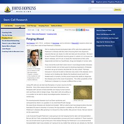
Dr. Current Opinion in Genetics & Development - Prospects for stem cell therapeutics: myths and medicines. Abstract With common scientific themes and experimental strategies, stem cell biology is evolving into a recognizable discipline.

Its clinical arm, regenerative medicine, is also gaining momentum—invigorated by the potential of stem cells to provide treatments for a host of medical conditions that are poorly served by drug therapy. But are the expectations for stem cell therapies realistic or overstated? In the past year, neurons, insulin-producing cells, and hematopoietic stem cells have been generated from embryonic stem cells or cultivated from somatic tissues of the adult. These cells have yielded modest and preliminary hints of functional reconstitution in animal models. Keywords embryonic stem cells; therapeutic cloning; plasticity; transdifferentiation; cell therapy; pluripotency Cell biology; Genetics; Development; Molecular Medicine. Neuron - Neurobiology of Depression. To view the full text, please login as a subscribed user or purchase a subscription.
Click here to view the full text on ScienceDirect. Figure 1. Behavioural Brain Research - The structural and functional connectivity of the amygdala: From normal emotion to pathological anxiety. Abstract The dynamic interactions between the amygdala and the medial prefrontal cortex (mPFC) are usefully conceptualized as a circuit that both allows us to react automatically to biologically relevant predictive stimuli as well as regulate these reactions when the situation calls for it.
In this review, we will begin by discussing the role of this amygdala–mPFC circuitry in the conditioning and extinction of aversive learning in animals. We will then relate these data to emotional regulation paradigms in humans. Finally, we will consider how these processes are compromised in normal and pathological anxiety. We conclude that the capacity for efficient crosstalk between the amygdala and the mPFC, which is represented as the strength of the amygdala–mPFC circuitry, is crucial to beneficial outcomes in terms of reported anxiety. Highlights. Www.if-pan.krakow.pl/pjp/pdf/2009/5_761.pdf. 2012 Program - Molecular & Cellular Neurobiology. The 2012 Gordon Research Conference on Molecular and Cellular Neurobiology will facilitate the interaction of global neuroscientists to demystify brain function and explicate the causes of brain disorders.
The meeting will bring together neuroscientists working at the forefront of the molecular and cellular basis of neuronal polarity, circuitry assembly and function, mechanisms of plasticity, molecular basis of behavior, regenerative neuroscience, and brain disorders, allow for in-depth discussion of most recent advances and stimulate the interdisciplinary and international collaboration.
The Conference will provide opportunities for junior scientists and graduate students to present their work in poster format and exchange ideas with leaders in the field. Some poster presenters will be selected for short talks. The project described was supported by Grant Number R13NS078948 from the National Institute Of Neurological Disorders And Stroke.