
THE AMYGDALA AND THE EMOTIONS. Chapter 9 — The Amygdala and the Emotions by Ben Best This installment is something of a digression in my "systematic" attempt to investigate the anatomical basis of mind.
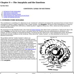
Here I investigate in detail a single component of the "Limbic System": the amygdala. The basis of this investigation is a book of scientific papers entitled THE AMYGDALA, Edited by John P. Aggleton (Wiley-Liss, 1992). Can mental activity protect against memory problems in multiple sclerosis? A new study shows that a mentally active lifestyle may protect against the memory and learning problems that often occur in multiple sclerosis (MS).
The study is published in the June 15, 2010, print issue of Neurology®, the medical journal of the American Academy of Neurology. "Many people with MS struggle with learning and memory problems. This study shows that a mentally active lifestyle might reduce the harmful effects of brain damage on learning and memory. That is, learning and memory ability remained quite good in people with enriching lifestyles, even if they had a lot of brain damage (brain atrophy on brain scans).
In contrast, persons with lesser mentally active lifestyles were more likely to suffer learning and memory problems, even at milder levels of brain damage," said study author James Sumowski, PhD, with the Kessler Foundation Research Center in West Orange, New Jersey. THE BRAIN FROM TOP TO BOTTOM. For many years, scientists’ understanding of how the brain processes language was rather simple: they believed that Wernicke’s area interpreted the words that we hear or read, then relayed this information via a dense bundle of fibres to Broca’s area, which generated any words that we spoke in response.
But subsequent experiments with brain imaging have revealed the existence of a third region of the brain that is also indispensable for language. This region is the inferior parietal lobule, also known as “Geschwind’s territory”, in honour of the American neurologist Norman Geschwind, who foresaw its importance as early as the 1960s. Brain imaging studies have now shown that the inferior parietal lobule (angular gyrus and supramarginal gyrus) is connected by large bundles of nerve fibres to both Broca’s area and Wernicke’s area. The Whole Brain Atlas.
Mini-strokes leave 'hidden' brain damage. Each year, approximately 150,000 Canadians have a transient ischemic attack (TIA), sometimes known as a mini-stroke.

New research published January 28 in Stroke, the journal of the American Heart Association shows these attacks may not be transient at all. They in fact create lasting damage to the brain. The stroke research team, led by Dr. Lara Boyd, physical therapist and neuroscientist with the Brain Research Centre at Vancouver Coastal Health and the University of British Columbia, studied 13 patients from the Stroke Prevention Clinic at Vancouver General Hospital and compared them against 13 healthy study participants. The TIA subjects had all experienced an acute episode affecting motor systems, but had symptoms resolved within 24 hours. "What we found has never been seen before," says Dr. "These findings are very important," says Dr.
Brain Basics - 3D Model of Brain Injury. Animated Deceleration Injury from a Traumatic Brain Injury TBI Inform: Introduction to Brain Injury What Happens When a Brain Bleeds?

Making New Connections: How the Brain Can Develop Into Adulthood - Alice G. Walton - Life. A multi-year study has found that the brain's white matter -- fibers connecting cells -- can shift with employment, education, and relationships For many years, the convention was that after adolescence, the brain had done about as much developing as it was going to do.
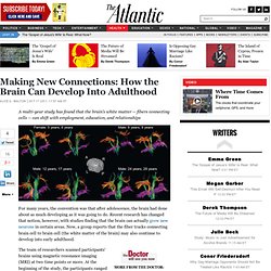
Recent research has changed that notion, however, with studies finding that the brain can actually grow new neurons in certain areas. Now, a group reports that the fiber tracks connecting brain cell to brain cell (the white matter of the brain) may also continue to develop into early adulthood. The team of researchers scanned participants' brains using magnetic resonance imaging (MRI) at two time points or more.
At the beginning of the study, the participants ranged in age from 5 to 29, and the average gap between the first and second scanning was about four years. The frontal lobe is responsible for high-level executive function and attention. This article originally appeared on TheDoctorWillSeeYouNow.com. BrainLine.org. EBP of TBI. Brain fitness.