Influenza. Typically, influenza is transmitted through the air by coughs or sneezes, creating aerosols containing the virus.
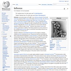
Influenza can also be transmitted by direct contact with bird droppings or nasal secretions, or through contact with contaminated surfaces. Airborne aerosols have been thought to cause most infections, although which means of transmission is most important is not absolutely clear.[4] Influenza viruses can be inactivated by sunlight, disinfectants and detergents.[5][6] As the virus can be inactivated by soap, frequent hand washing reduces the risk of infection.[7] Flu can occasionally lead to pneumonia, either direct viral pneumonia or secondary bacterial pneumonia, even for persons who are usually very healthy.[8][9][10] In particular it is a warning sign if a child (or presumably an adult) seems to be getting better and then relapses with a high fever as this relapse may be bacterial pneumonia.[11] Another warning sign is if the person starts to have trouble breathing.[10]
Epithelium. Epithelial layers are avascular, so they must receive nourishment via diffusion of substances from the underlying connective tissue, through the basement membrane.[2][3] Epithelia can also be organised into clusters of cells that function as exocrine and endocrine glands.
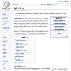
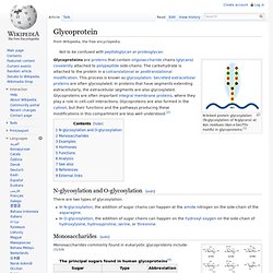
Classification[edit] Summary showing different epithelial cells/tissues and their characteristics. There are three principal morphologies associated with epithelial cells: Squamous epithelium has cells that are wider than they are tall (flat and scale-like).Cuboidal epithelium has cells whose height and width are approximately the same (cube shaped).Columnar epithelium has cells taller than they are wide (column-shaped). Sialic acid. Sialic acid is a generic term for the N- or O-substituted derivatives of neuraminic acid, a monosaccharide with a nine-carbon backbone.[1] It is also the name for the most common member of this group, N-acetylneuraminic acid (Neu5Ac or NANA).
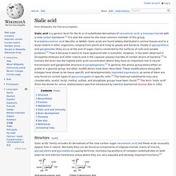
Sialic acids are found widely distributed in animal tissues and to a lesser extent in other organisms, ranging from plants and fungi to yeasts and bacteria, mostly in glycoproteins and gangliosides (they occur at the end of sugar chains connected to the surfaces of cells and soluble proteins).[2] That is because it seems to have appeared late in evolution. Structure[edit] Sialic acids' family includes 43 derivatives of the nine-carbon sugar neuraminic acid, but these acids unusually appear free in nature. The numbering of the sialic acid structure begins at the carboxylate carbon and continues around the chain. The configuration that places the carboxylate in the axial position is the alpha-anomer. Glycoprotein. N-linked protein glycosylation (N-glycosylation of N-glycans) at Asn residues (Asn-x-Ser/Thr motifs) in glycoproteins.[1] N-glycosylation and O-glycosylation[edit] There are two types of glycosylation: In N-glycosylation, the addition of sugar chains can happen at the amide nitrogen on the side-chain of the asparagine.In O-glycosylation, the addition of sugar chains can happen on the hydroxyl oxygen on the side-chain of hydroxylysine, hydroxyproline, serine, or threonine.
Monosaccharides[edit] Eight sugars commonly found in glycoproteins. Monosaccharides commonly found in eukaryotic glycoproteins include:[3]:526 The sugar group(s) can assist in protein folding or improve proteins' stability. Endocytosis. The different types of endocytosis Endocytosis pathways[edit] Endocytosis pathways could be subdivided into four categories: namely, clathrin-mediated endocytosis, caveolae, macropinocytosis, and phagocytosis.[2] More recent experiments have suggested that these morphological descriptions of endocytic events may be inadequate, and a more appropriate method of classification may be based upon the clathrin-dependence of particular pathways, with multiple subtypes of clathrin-dependent and clathrin-independent endocytosis.
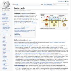
Mechanistic insight into non-phagocytic, clathrin-independent endocytosis has been lacking, but a recent study has shown how Graf1 regulates a highly prevalent clathrin-independent endocytic pathway known as the CLIC/GEEC pathway.[5] Amantadine. Amantadine (trade name Symmetrel, by Endo Pharmaceuticals) is a drug that has U.S.
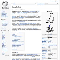
Food and Drug Administration approval for use both as an antiviral and an antiparkinsonian drug. It is the organic compound 1-adamantylamine or 1-aminoadamantane, meaning it consists of an adamantane backbone that has an amino group substituted at one of the four methyne positions. Rimantadine is a closely related derivative of adamantane with similar biological properties.
Apart from medical uses, this compound is useful as a building block, allowing the insertion of an adamantyl group. According to the U.S. History[edit] Amantadine was approved by the U.S. Aciclovir. Aciclovir (INN, BAN.
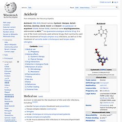
Brand names: Cyclovir, Herpex, Acivir, Acivirax, Zovirax, Zoral, Xovir and Imavir) /eɪˈsaɪklɵvɪər/ or acyclovir (USAN, former BAN), chemical name acycloguanosine, abbreviated as ACV,[2] is a guanosine analogue antiviral drug. Gene therapy. Gene therapy using an adenovirus vector. A new gene is inserted into a cell using an adenovirus. If the treatment is successful, the new gene will make functional protein to treat a disease.
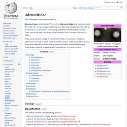
Gene therapy was first conceptualized in 1972, with the authors urging caution before commencing gene therapy studies in humans. Adenoviridae. They have a broad range of vertebrate hosts; in humans, 57 distinct adenoviral serotypes have been found to cause a wide range of illnesses, from mild respiratory infections in young children to life-threatening multi-organ disease in people with a weakened immune system.
Golgi apparatus. Micrograph of Golgi apparatus, visible as a stack of semicircular black rings near the bottom.
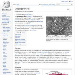
Numerous circular vesicles can be seen in proximity to the organelle. Part of the cellular endomembrane system, the Golgi apparatus packages proteins inside the cell before they are sent to their destination; it is particularly important in the processing of proteins for secretion. Discovery Owing to its large size, the Golgi apparatus was one of the first organelles to be discovered and observed in detail. It was discovered in 1898 by Italian physician Camillo Golgi during an investigation of the nervous system.[2] After first observing it under his microscope, he termed the structure the internal reticular apparatus.