
Electron Microscopy Lab. Dedicated to the characterization of materials through imaging, chemical, and crystallographic analyses of material microstructures in support of Basic Energy Science, Laboratory Directed Research and Development, DoD, DOE, Work for Others, nuclear energy, and weapons programs.
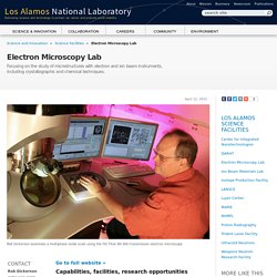
Go to full website » Capabilities, facilities, research opportunities The Electron Microscopy Lab (EML) is a user facility maintained by the Lab's Metallurgy Group (MST-6) of the Materials Science Division. Light Sheet Microscopy. Casos clinicos. Microscope. Histologie. Steve Chapman SEM & TEM Courses World Wide. Steve Chapman formed Protrain in 1982 and he is our senior consultant in electron microscopy.
Steve first became involved with electron microscopes in 1964 at the University of Birmingham, England. Moving on to working as an EM service engineer in the UK his service activities as a senior engineer eventually took him throughout Europe. He then moved into sales and marketing where he worked with a number of electron microscope and accessory manufacturers in the demonstration and application fields. Steve has been involved with the design of cryo systems and sputter coaters for SEM, as well as the design of transmission and scanning electron microscopes themselves. Running courses in many parts of the world, north and south of the equator, he has written six books including those on the operation and maintenance of scanning and transmission electron microscopes.
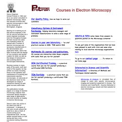
Steve is also very active in his other love that of motorsport. Our Quality Policy, how we hope to serve our customers. Electron Microscopy: Freezing Samples. Graphics courtesy of IDG Books Cryo EM is a microscopy technique in which the sample to be viewed is frozen in a very cold liquid refrigerant in order to preserve and protect it during observation.
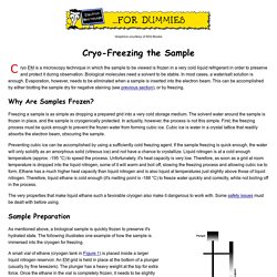
Biological molecules need a solvent to be stable. In most cases, a water/salt solution is enough. Evaporation, however, needs to be eliminated when a sample is inserted into the electron beam. This can be accomplished by either blotting the sample dry for negative staining (see previous section), or by freezing. The Life and Death of a Tungsten Hairpin Filament. Many words have been written about filament saturation and filament life, not all with years of experience behind them!
Sometimes when visiting laboratories one has the feeling that the life of the filament is far more important than the quality of the results, so let us try to demystify filament life? The life of a tungsten hairpin filament very much depends upon its use, the applications in a particular laboratory. Medium to low magnification applications in the transmission electron microscope (TEM) may only require low emission currents (10 to 15 micro amps) with a long filament life resulting. High resolution studies require higher operating currents (20 to 45 micro amps) and when demanding more emission the life of the filament will suffer. Www.santoshraut.com. Microscopy: Microscopy is any technique for producing visible images of structures or details too small to otherwise be seen by the human eye, using a microscope or other magnification tool.
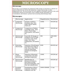
The microscope was first applied to biological material in the early seventeenth century by researchers such as Robert Hooke and Antoni van Leeuwenhoek, and remains a pivotal tool in learning about biology today. Brief Introduction to Freeze Fracture and Etching: Leica Science Lab. Fit for the environment of an electron microscopeSublimation and condensation of water – Freeze etching and contaminationFreeze fracture to generate imagesUnidirectional shadowing of freeze fractured yeastApplicationsDownload Fig. 1: Plant-louse on a wheat leave The chamber of an electron microscope is evacuated to a very low pressure.
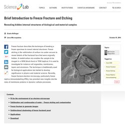
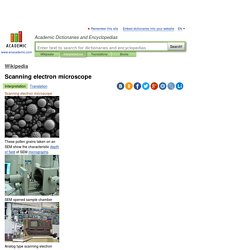
The structure of a living cell placed into this environment cannot be preserved due to the extremely quick evaporation of the water which makes most part of the cell. Methodology at University of Maryland - Baltimore County - StudyBlue. Scanning electron microscope. SEMopenedsamplechamber Analogtypescanningelectronmicroscope.
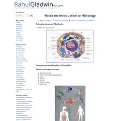
Rahul's Noteblog: Introduction to Histology. Rahul's Noteblog Notes on Histology Notes on Introduction to Histology.
SPIDER: WWW Sites for Biological Electron Microscopy. History of Biology: Cell Theory and Cell Structure - Biology Encyclopedia - cells, plant, body, function, process, animal, different, organisms, chromosomes. Photo by: Russi & Morelli All living organisms are composed of cells, and all cells arise from other cells.
These simple and powerful statements form the basis of the cell theory, first formulated by a group of European biologists in the mid-1800s. So fundamental are these ideas to biology that it is easy to forget they were not always thought to be true. Robert Hooke's microscope. Hooke first described cells in 1665. Early Observations. Histology-World! Histology. Home Page of the Electron Microscopy Unit and Jokitalo group. New & Used SEM's, TEM's For Sale. Microscopy Production & Sales. Companies - Electron microscopy.
Showing company 1 to 20 of 74 in Electron microscopy.
Result page: 1 2 3 4 Next » XEI Scientific, Inc.May 21, 2014 XEI Scientific Inc. invented the Evactron De-Contaminator in 1999 as the first plasma cleaner to use a downstream cleaning process to remove carbon from electron microscopes. M.GLOBALSOURCES.COM. The MicroscopeMaster Blog. Best Microscope Reviews and Microscopy Research. The SPI Supplies List of Blogs for Microscopy and Microanalysis. "Blogging" or the act of maintaining a "blog" is the fastest growing part of the "internet community". To some degree, blogs mean different things to different people, and we have seem some that were labeled as "blogs" but were more just newsletters or periodic mailings. Some call this part of the electronic media "Web 2.0".
We like to think of a blog as a running commentary of an individual or group of persons, who make periodic postings on some topic of interest along with the provision for others who read their blog postings, to comment and for yet others to comment on the comments. Some consider the following of blogs to be a waste of time. On the other hand, there are those who have followed some of the investment advice (e.g. The Microscopy Blog - Microscope Experiments, Techniques, and Miscellany. Microscope World Blog. Society of America President's Blog. Electron microscope photography - Microscopy Blog.
Imaging & Microscopy Resource. The Arduino IDE for ARM microcontrollers is just plain awesome. I’ve been using both an Uno and a Mega board for quite a while now, and continue to be impressed with how flexible these inexpensive boards are. Recently, I wanted to read the output of a pair of rotary encoders, like these: Encoders can be used for all sorts of things, from acting as a user control knob (with endless turns), to acting as a verification system for motors, to confirm that a commanded # of motor moves has been completed accurately. In fact, linear and rotary encoders are used on microscope stages for this exact reason, to confirm that the stage actually went some specified distance.
So – I wanted to use an encoder as a knob for user input. Encoders output a set of signals, which run either open or closed against a ground, based on the position and direction of the knob turn. Byte range = 50; if ( positionLeft > range){ knobLeft.write(range); } if ( positionLeft < -range){ knobLeft.write(-range); } MicrobeHunter.com - Microscopy Magazine and Blog. Thoughts on imaging from the director of the NIC@UCSF/QB3. I just came across this interesting PDF summarizing the performance of Point Grey cameras. Point Grey is a machine vision camera manufacturer, and I don’t normally think of using machine vision cameras for microscopy. Directory of Electron Microscopy Facilities in Canada. Electron Microscopy and Microanalysis, Carleton University Research Facility for (CURFEMM) (CNS) - Directories.
Microscopy Imaging Center. Imaging in the Biomedical and Materials Sciences The Microscopy Imaging Center (MIC) is a College of Medicine Core Facility designed as a multi-user resource for sample preparation and collection and analysis of images for biological and materials applications. Additionally, the MIC is a CAP certified laboratory performing electron microscopic clinical diagnosis. Marine Biological Laboratory. This course is designed primarily for research scientists, postdoctoral trainees, and advanced graduate students in animal, plant, medical, and material sciences. Non-biologists seeking a comprehensive introduction to microscopy and digital imaging will benefit greatly from the course. Some prior theoretical or practical understanding of the basic principles of optics and microscopy is necessary. This 10 day course is limited to 26 students. Microscopy and Analysis. Microscopy and Analysis. MML What We Do. IntroductionImportancePeople and Facilities Introduction.
Micrographia: A Light Microscopy Resource: Home Page and Site Directory. Science: Methods and Techniques: Microscopy: Light Microscopy. Index. Directory of Microscopy and Microanalysis WWW Sites. Microscopy-News.com. News Microscopy. EMBO Practical Course - Cryo-Electron Microscopy and 3D Image Processing - 26 August - 2 September 2012. Microscopy-News.com - Microscopy-News.com - Portal for Press and Products. Microscopy Today Online - Home. MICROSCOPIA ELECTRÓNICA. A diferença básica entre as duas modalidades de microscopia, consiste no facto de na primeira, a imagem ser produzida por fotões e na segunda, por electrões. Daí decorrem necessariamente, consequências, quer a nível de concepção física dos aparelhos, quer a nível da preparação do material, quer a nível da natureza da imagem e ou ainda da performance da técnica. Microscópio electrónico de transmissão (Transmission electron microscope) O microscópio electrónico de transmissão é habitualmente designado abreviadamente pelas iniciais doseu nome em inglês: TEM.
Desenvolveu-se nos anos 30. Tal como no microscópio fotónico, também no microscópio electrónico é composto por uma fonte (de electrões), um sistema ocular, um sistema objectiva e um sistema condensador. A fonte consiste num filamento metálico tornado incandescente pelo efeito de Joule. Ao atravessar a preparação, os electrões são desviados uns mais do que outros. Poder de resolução l = h / mv Ampliação Preparação dos espécimes Fixação. Monocomp. Page K2: Sputter Coaters & Carbon Evaporators - Turbo Pumped - ProSciTech Pty. Ltd. HIGH VACUUM EVAPORATOR for Multiple Applications - EMITECH K975XThe K975X Turbo Evaporator, is a multiple application system to enable a range of preparation techniques to be applied with the flexibility and module expansion capability to develop new methods and prepare new specimens.
Histology. Repair for microtomes, ultramicrotomes, tissue processors, vibratomes, plunge freezers, ovens, baths, shakers, freeze fracture, vacuum systems.